

Acute dacryocystitis with giant lacrimal abscess: a case report
- PMID: 32014022
- PMCID: PMC6998065
- DOI: 10.1186/s13052-020-0779-7
Acute dacryocystitis with giant lacrimal abscess: a case report
Abstract
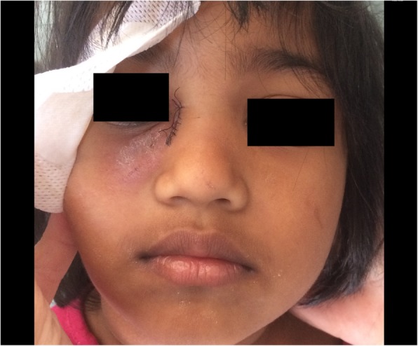
Background: We report a case of a 4-year-old girl with acute dacryocystitis complicated with giant lacrimal abscess who underwent open dacryocystectomy as resolutive surgery.
Case presentation: A 4-year-old previously healthy girl presented to the emergency department with a voluminous and erythematous, fluctuant warm mass localized inferiorly to the medial canthus of the right eye. She had a 2-week history of right inferior eyelid oedema and hyperemia, treated firstly with dexamethasone and netilmicin by eye drops, and then with per oral amoxicillin clavulanate. Ultrasound examination showed a well-circumscribed round lesion filled by anechoic fluid with punctate echoes, confirming a diagnosis of acute dacryocystitis complicated by lacrimal abscess. Parents refused a head CT. Systemic antibiotic treatment was started and, on 5th day from admission, open dacryocystectomy was performed with good esthetical result.
Conclusions: Pediatric acute dacryocystitis is a potentially serious condition, which must be treated with intravenous antibiotic therapy followed by surgery tailored to the clinical history. Even if probing and dacryocystorhinostomy are the most used surgery in adults and children, open dacryocystectomy is a safe and successful option, mainly in severe cases where imaging studies are not available.
Keywords: Dacryocystectomy; Dacryocystorhinostomy; Lacrimal abscess; Nasolacrimal duct obstruction; Pediatric acute dacryocystitis.
Conflict of interest statement
The authors declare that they have no competing interests.
Figures

References
-
- Campolattaro BN, Lueder GT, Tychsen L. Spectrum of pediatric dacryocystitis: medical and surgical management of 54 cases. J Pediatr Ophthalmol Strabismus. 1997;34:143–153. - PubMed
Publication types
MeSH terms
Substances
LinkOut - more resources
Full Text Sources
Medical
