Review
. 2020 Feb 3;15(1):39.
doi: 10.1186/s13023-020-1320-1.

Kaposiform hemangioendothelioma: current knowledge and future perspectives

Affiliations
Review

Kaposiform hemangioendothelioma: current knowledge and future perspectives

Yi Ji et al. Orphanet J Rare Dis. .

Abstract

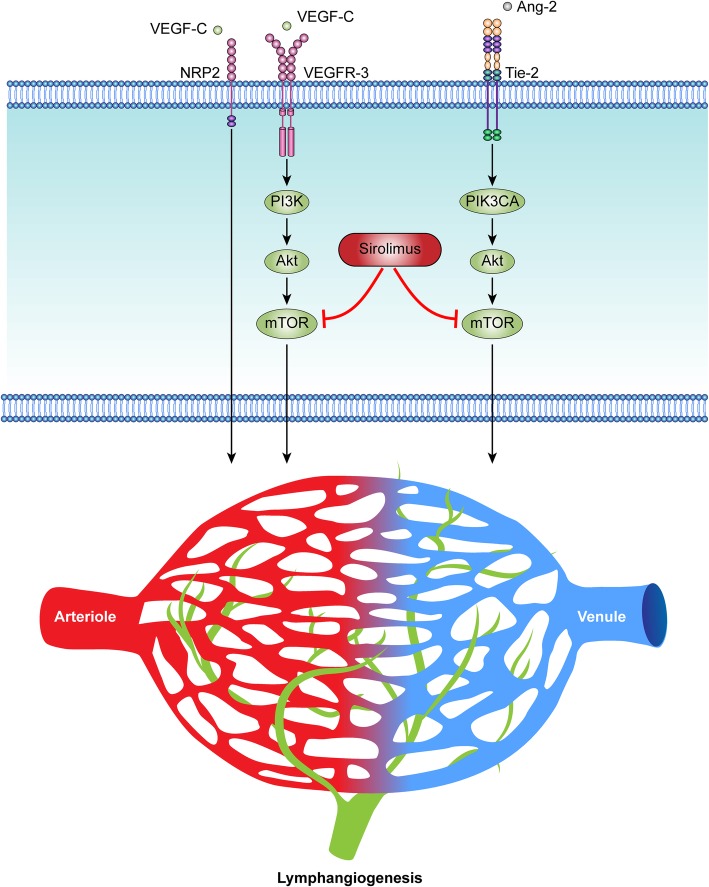
Kaposiform hemangioendothelioma (KHE) is a rare vascular neoplasm with high morbidity and mortality. The initiating mechanism during the pathogenesis of KHE has yet to be discovered. The main pathological features of KHE are abnormal angiogenesis and lymphangiogenesis. KHEs are clinically heterogeneous and may develop into a life-threatening thrombocytopenia and consumptive coagulopathy, known as the Kasabach-Merritt phenomenon (KMP). The heterogeneity and the highly frequent occurrence of disease-related comorbidities make the management of KHE challenging. Currently, there are no medications approved by the FDA for the treatment of KHE. Multiple treatment regimens have been used with varying success, and new clinical trials are in progress. In severe patients, multiple agents with variable adjuvant therapies are given in sequence or in combination. Recent studies have demonstrated a satisfactory efficacy of sirolimus, an inhibitor of mammalian target of rapamycin, in the treatment of KHE. Novel targeted treatments based on a better understanding of the pathogenesis of KHE are needed to maximize patient outcomes and quality of life. This review summarizes the epidemiology, etiology, pathophysiology, clinical features, diagnosis and treatments of KHE. Recent new concepts and future perspectives for KHE will also be discussed.

Keywords: Angiogenesis; Kaposiform hemangioendothelioma; Kasabach-Merritt phenomenon; Lymphangiogenesis; Treatment.


Conflict of interest statement

The authors declare that they have no competing interests, either financial or non-financial, that could be perceived as prejudicing the impartiality of the research reported.

Figures

Fig. 1
Fig. 1
A 3-month-old male infant presenting KHE associated with KMP. The tumor lesion grew progressively after the first week of age and extended through the whole thigh, scrotum and abdominal wall, with ‘extensive thrombocytopenic purpura’ as described by Kasabach and Merritt in 1940
Fig. 2
Fig. 2
G-protein-coupled receptors (GPCRs) participate in different physiological processes. The binding of ligands to GPCRs triggers a universal G protein allosteric mechanism that promotes the exchange of GDP with GTP on the α subunit of heterotrimeric G proteins. This event causes the dissociation of Gα from the dimer. Gα subunits mediate signals between GPCRs and intracellular signaling cascades. These signaling pathways include the PI3K/AKT/mTOR and MAPK/ERK pathways, both of which can mediate different biological processes, such as cell proliferation, migration and survival. Mutations in GPCRs and G proteins have been found at a very high frequency in tumor cells and endothelial cells in vascular anomalies
Fig. 3
Fig. 3
VEGF-C/VEGFR3 and Ang-2/Tie-2 signaling pathways play an important role in lymphangiogenesis. The binding of VEGF-C can stimulate the activation of VEGFR-3 and induce downstream PI3K/Akt/mTOR signaling, which mediates lymphangiogenesis. VEGF-C binding to NRP-2 can form a complex with VEGFR-3, further activating the VEGFR-3 signaling that enhances the proliferation of lymphatic endothelial cells (LECs) and lymphangiogenesis. Ang-2 ligand-induced Tie-2 activation triggers Akt/mTOR signaling, which in LECs is mainly mediated by PIK3CA. Gene-targeting experiments have identified that the Ang-2/Tie-2 signaling system is needed for physiological and pathological remodeling of lymphatic vessels. Sirolimus (rapamycin), which is an inhibitor of mTOR, complements current antilymphatic strategies in the treatment of vascular anomalies, such as KHE
Fig. 4
Fig. 4
Deep KHE with bone and joint destruction in a 3.5-year-old girl. The patient had been diagnosed with a left-hip KHE associated with KMP at 1 year of age. She received a partial resection at the local hospital. Although surgical excision improved the associated KMP, she exhibited a progressively decreased hip range of motion. An anteroposterior pelvis radiograph showed right hip subluxation and progressive bone erosion in the left ilium and proximal femur before referral (a, b and c). Coronal T2-weighted MRI revealed a deep lesion infiltrating the left ilium and proximal femur at the time of referral to our department (d, e)
Fig. 5
Fig. 5
Clinical and MRI features of KHE with KMP. a A 2-month-old boy was found to have a chest wall mass after birth. The mass became progressively indurated and purpuric. The boy developed profound thrombocytopenia and consumptive coagulopathy. b Horizontal T1-weighted MRI revealed that the heterogeneous mass was isointense relative to the adjacent muscle on T1-weighted imaging. Horizontal (c) and sagittal (d) T2-weighted MRI revealed hyperintense lesions infiltrating the right lateral chest wall
Fig. 6
Fig. 6
Histopathological characteristics of KHE. aHematoxylin and eosin (H&E)-stained sections of KHE (original magnification × 100). The histologic hallmark of KHE was infiltrating, defined, rounded and confluent nodules, which were composed of spindle endothelial cells. b These spindle endothelial cells aligned to form malformed lymphatic channels and a slit-like vascular lumina (× 200). Immunohistochemical staining showed that the endothelial cells in KHE lesions were positive for the vascular endothelial markers CD31 (c, × 100) and CD34 (d, × 100) and the lymphatic endothelial marker D2–40 (e, × 100) but were negative for glucose transporter-1 and human herpes virus-8 staining (data not shown). Ki-67 was noted in only a few nuclei in the lesion (f, × 100)
Fig. 7
Fig. 7
Rapidly involuting congenital hemangiomas (RICH). a RICH is fully formed at birth and then involutes, mostly during the first year of life. The patient’s hematologic parameters were within the normal reference ranges. b The same RICH involuted rapidly without any treatment. By 9 months of age, the lesion had involuted completely, leaving dermal atrophy
Fig. 8
Fig. 8
Congenital KHE associated with KMP on the right face. Congenital KHEs with KMP likely represent a period of temporary and partial remission shortly after birth. The signs and symptoms may alleviate spontaneously. However, rebound growth of the lesions accompanying severe KMP would reoccur within the next several days or weeks. a The parents’ photograph revealed a bluish, swollen and firm vascular mass on the right face after birth (2 days of age). Her platelet count was 7 × 109/L. Without any special treatment, the tumor became soft and was stagnant in size. Her platelet count reached a highest value of 161 × 109/L (b: 1 week of age, c: 6.5 weeks of age). Subsequently, however, the tumor became progressively enlarged and displayed obvious ecchymosis (d: 8 weeks of age). The patient’s platelet count dropped to 3 × 109/L. She was treated with a combination therapy of sirolimus (0.8 mg/m2 administered twice daily) and prednisolone (2 mg/kg/d). One (e) and 4 weeks (f) after treatment. Within 10 days of combination therapy, the girl’s platelet level normalized. The prednisolone was tapered and discontinued within the following 4 weeks, and sirolimus was continued. G, Twelve months after treatment. H, Photograph at 26 months of age (24 months of treatment) demonstrates a nearly complete involution of the lesion. Sirolimus was then tapered and discontinued

Similar articles

Cited by

References

    1. O'Rafferty C, O'Regan GM, Irvine AD, Smith OP. Recent advances in the pathobiology and management of Kasabach-Merritt phenomenon. Brit J Haematol. 2015;171(1):38–51. doi: 10.1111/bjh.13557. - DOI - PubMed
    1. Lyons LL, North PE, Mac-Moune LF, Stoler MH, Folpe AL, Weiss SW. Kaposiform hemangioendothelioma: a study of 33 cases emphasizing its pathologic, immunophenotypic, and biologic uniqueness from juvenile hemangioma. Am J Surg Pathol. 2004;28(5):559–568. doi: 10.1097/00000478-200405000-00001. - DOI - PubMed
    1. Mulliken JB, Anupindi S, Ezekowitz RAB, Mihm MC. Case 13-2004. New Engl J Med. 2004;350(17):1764–1775. doi: 10.1056/NEJMcpc049002. - DOI - PubMed
    1. Schaefer BA, Wang D, Merrow AC, Dickie BH, Adams DM. Long-term outcome for kaposiform hemangioendothelioma: a report of two cases. Pediatr Blood Cancer. 2017;64(2):284–286. doi: 10.1002/pbc.26224. - DOI - PubMed
    1. Fernandez-Pineda I, Lopez-Gutierrez JC, Chocarro G, Bernabeu-Wittel J, Ramirez-Villar GL. Long-term outcome of vincristine-aspirin-ticlopidine (VAT) therapy for vascular tumors associated with kasabach-Merritt phenomenon. Pediatr Blood Cancer. 2013;60(9):1478–1481. doi: 10.1002/pbc.24543. - DOI - PubMed

Publication types

Supplementary concepts