. 2020 Feb 3;8(1):10.
doi: 10.1186/s40478-020-0889-x.

Genetic and histologic spatiotemporal evolution of recurrent, multifocal, multicentric and metastatic glioblastoma

Affiliations

Genetic and histologic spatiotemporal evolution of recurrent, multifocal, multicentric and metastatic glioblastoma

Maria-Magdalena Georgescu et al. Acta Neuropathol Commun. .

Abstract

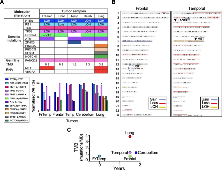
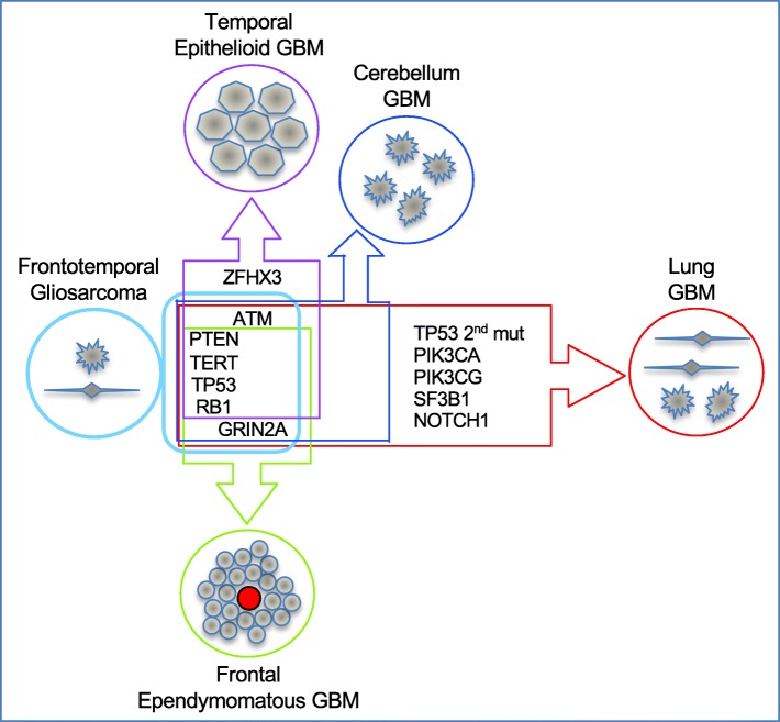
Glioblastoma is the most frequent and aggressive primary brain tumor, characterized by extensive brain invasion and rarely, systemic metastases. The pathogenesis of metastatic glioblastoma is largely unknown. We present the first integrated clinical/histologic/genetic analysis of 5 distinct brain and lung foci from a unique case of recurrent, multifocal, multicentric and metastatic glioblastoma. The initial right frontotemporal gliosarcoma received standard surgical/chemoradiation therapy and recurred 1.5 years later, co-occurring with three additional masses localized to the ipsilateral temporal lobe, cerebellum and lung. Synchronous metastatic lung carcinoma was suspected in this long-term smoker patient with family history of cancer. However, glioblastoma was confirmed in all tumors, although with different morphologic patterns, including ependymomatous and epithelioid. Genomic profiling revealed a germline FANCD2 variant of unknown significance, and a 4-gene somatic mutation signature shared by all tumors, consisting of TERT promoter and PTEN, RB1 and TP53 tumor suppressor mutations. Additional GRIN2A and ATM heterozygous mutations were selected in the cerebellar and lung foci, but were variably present in the supratentorial foci, indicating reduced post-therapeutic genetic evolution in brain foci despite morphologic variability. Significant genetic drift characterized the lung metastasis, likely explaining the known resistance of circulating glioblastoma cells to systemic seeding. MET overexpression was detected in the initial gliosarcoma and lung metastasis, possibly contributing to invasiveness. This comprehensive analysis sheds light on the temporospatial evolution of glioblastoma and underscores the importance of genetic testing for diagnosis and personalized therapy.

Keywords: Epithelioid; Gliosarcoma; Invasion; Metastatic glioblastoma; Multicentric; Multifocal; Next generation sequencing; PTEN; Recurrent.


Conflict of interest statement

The authors declare that they have no competing interests.

Figures

Fig. 1
Fig. 1
Clinical case presentation. a Time course of the patient’s clinical progression: the initial right frontotemporal (FrTemp) mass was surgically removed and the patient received standard radiotherapy (XRT) and temozolomide (TMZ). At 18 months postresection, 4 new ipsilateral masses occurred: frontal (Fr), temporal (Temp), as well as in the lung and cerebellum near midline, and underwent resections, followed by chemotherapy. b. MRI at the time of recurrence, 18 months following the 1st craniotomy for the initial mass resection. Serial axial T1W post-contrast (T1W-C) images show a multifocal mass comprising a frontal mass (Frontal, green arrows) at the site of the previous resection, and a temporal mass (Temporal, purple arrows). c Coronal T1W post-contrast (T1W-C) images showing rapid growth of a cerebellar mass (blue arrows) between the 2nd and the 3rd craniotomies. d Chest CT with contrast showing a solid mass in the right lung (red arrow)
Fig. 2
Fig. 2
Morphologic variability of glioblastoma diagnosed in all five tumors. a H&E, GFAP immunolabeling and reticulin special staining show the biphasic pattern of gliosarcoma in the initial frontotemporal mass: the glial component (right upper corner) expresses GFAP and lacks pericellular reticulin deposition, whereas the sarcomatous component (left lower corner) shows opposite phenotype. b Ependymomatous and epithelioid morphology of the frontal and temporal recurrent tumors from the 2nd resection, respectively. Note opposite GFAP labeling patterns with perivascular tumor cell arrangement in the frontal mass (vessels indicated with green arrows), NHERF1 dot-like microlumen labeling in the frontal mass and membranous staining in the temporal mass, high Ki-67 proliferation in the frontal mass, and very focal Cam5.2 cytokeratin staining in the temporal mass. c Resection H&E of the cerebellar mass shows a homogenous glial component, calcifications (black arrow) and damaged vessels obstructed by fibrin thrombi and with invasion of neoplastic cells (blue arrow). d The lung biopsy H&E shows fibrillary areas (inset) with necrosis (black arrow), and pleomorphic areas with brisk mitotic activity (red arrow) and high Ki-67 proliferation index. GFAP diffusely labels the entire tumor
Fig. 3
Fig. 3
Genomic profiling. a Summary table of genomic and transcriptomic analysis, and bar graph of somatic mutation composition and VAF. Color-coded shading indicates mutant variants, whereas white rectangles correspond to wild-type variants. RNA overexpression is shown by red rectangles. The VAF of individual mutations was normalized to the highest VAF in a given sample, that was arbitrarily set to 100%, due to LOH (see Material and methods). FrTemp, frontotemporal (initial mass); Front, frontal; Temp, temporal; Cereb, cerebellum; Loss, homozygous CN loss; #, additional TP53 mutation in the lung specimen. b Chromosomal CN array from the adjacent frontal and temporal recurrences showing common chromosomal alterations but also divergence of the temporal tumor by extensive chromosome losses. Both tumors have loss of chromosome 10, and the frontal mass has gain of chromosome 7, whereas the temporal mass has a more complex CN gain and LOH of chromosome 7. The RB1 locus with homozygous CN loss is encircled, and the loci for FANCD2 and MET are indicated by arrowheads. c TMB representation showing significant mutation accumulation in the lung mass
Fig. 4
Fig. 4
Genetic and morphologic spatiotemporal evolution model. Tumors are schematically shown as circles filled with cells of various morphologies. The initial gliosarcoma is shown in light blue color on the left, adjacent to a box representing its genetic composition of core clonal mutations on the left, and subclonal mutations on the right. The genetic drift is shown on the vertical axis for the closely related intraneural secondary/recurrent foci, and on the horizontal axis for the more divergent lung metastasis. Additional mutations are indicated in the corresponding tumor boxes, whereas the 4-gene core signature is common to all tumors. GBM, glioblastoma

References

    1. Beauchesne P. Extra-neural metastases of malignant gliomas: myth or reality? Cancers (Basel) 2011;3:461–477. doi: 10.3390/cancers3010461. - DOI - PMC - PubMed
    1. Beaumont TL, Kupsky WJ, Barger GR, Sloan AE. Gliosarcoma with multiple extracranial metastases: case report and review of the literature. J Neuro-Oncol. 2007;83:39–46. doi: 10.1007/s11060-006-9295-x. - DOI - PubMed
    1. Boisvert RA, Howlett NG. The Fanconi anemia ID2 complex: dueling saxes at the crossroads. Cell Cycle. 2014;13:2999–3015. doi: 10.4161/15384101.2014.956475. - DOI - PMC - PubMed
    1. Chen CC, Taniguchi T, D'Andrea A. The Fanconi anemia (FA) pathway confers glioma resistance to DNA alkylating agents. J Mol Med (Berl) 2007;85:497–509. doi: 10.1007/s00109-006-0153-2. - DOI - PubMed
    1. Eckerich C, Zapf S, Fillbrandt R, Loges S, Westphal M, Lamszus K. Hypoxia can induce c-Met expression in glioma cells and enhance SF/HGF-induced cell migration. Int J Cancer. 2007;121:276–283. doi: 10.1002/ijc.22679. - DOI - PubMed

Publication types