Comparative Study
. 2020 Apr 1;70(2):160-169.
doi: 10.30802/AALAS-CM-19-000035. Epub 2020 Feb 3.

Comparative Analysis of Cellular Immune Responses in Conventional and SPF Olive Baboons (Papio anubis)

Affiliations
Comparative Study

Comparative Analysis of Cellular Immune Responses in Conventional and SPF Olive Baboons (Papio anubis)

Elizabeth R Magden et al. Comp Med. .

Abstract

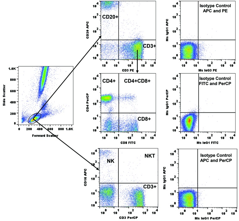
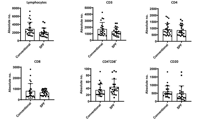
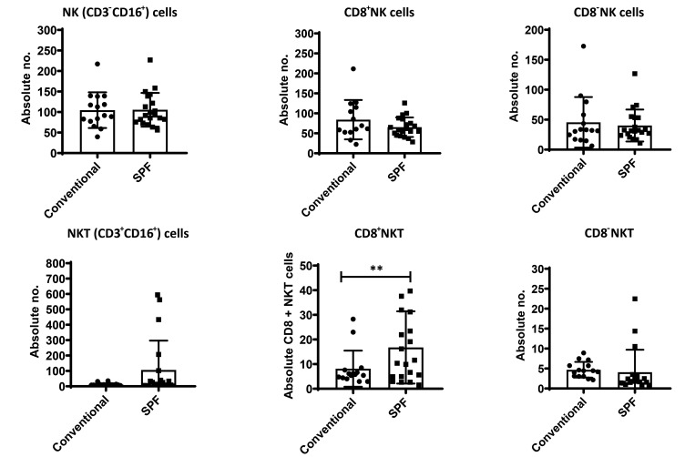
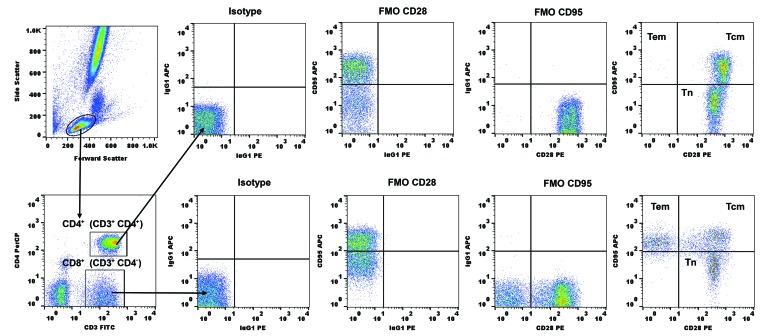
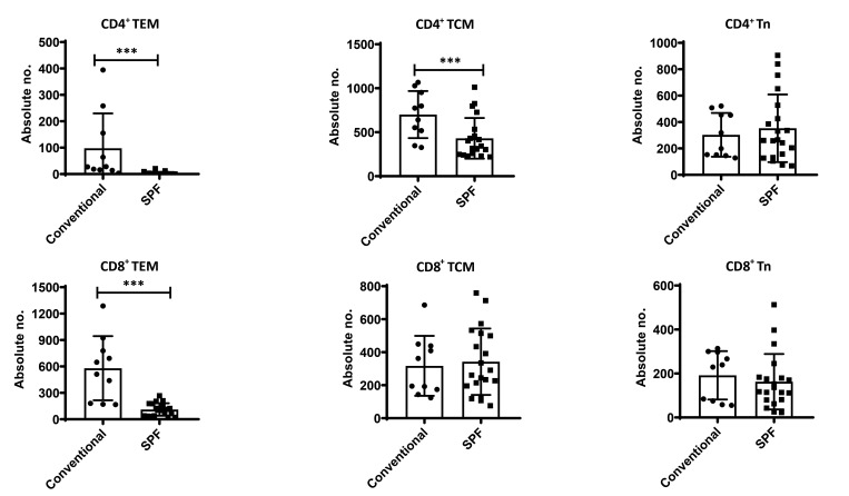
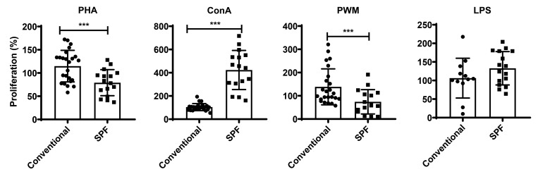
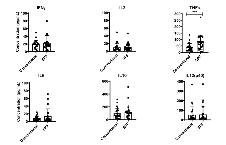
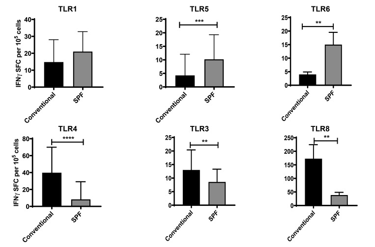
Olive baboons (P. anubis) have provided a useful model of human diseases and conditions, including cardiac, respiratory, and infectious diseases; diabetes; and involving genetics, immunology, aging, and xenotransplantation. The development of a immunologically defined SPF baboons has advanced research further, especially for studies involving the immune system and immunosuppression. In this study, we compare normal immunologic changes of PBMC subsets, and their function in age-matched conventional and SPF baboons. Our results revealed that both groups have comparable numbers of different lymphocyte subsets, but phenotypic differences in central and effector memory T-cell subsets are more pronounced in CD4+ T cells. Despite equal proportions of CD3+ T cells among the conventional and SPF baboons, PBMC from the conventional group showed greater proliferative responses to phytohemagglutinin and pokeweed mitogen and higher numbers of IFNγ-producing cells after stimulation with concanavalin A or pokeweed mitogen, whereas plasma levels of the inflammatory cytokine TNFα were significantly higher in SPF baboons. Exposure of PBMC from conventional baboons to various Toll-like (TLR) ligands, including TLR3, TLR4, and TLR8, yielded increased numbers of IFNγ producing cells, whereas PBMC from SPF baboons stimulated with TLR5 or TLR6 ligand had more IFNγ-producing cells. These findings suggest that although lymphocyte subsets share many phenotypic and functional similarities in conventional and SPF baboons, specific differences in the immune function of lymphocytes could differentially influence the quality and quantity of their innate and adaptive immune responses. These differences should be considered in interpreting experimental outcomes, specifically in studies measuring immunologic endpoints.


Figures

Figure 1.
Figure 1.
Gating scheme for phenotypic analyses of the various cell markers in peripheral blood from a representative baboon. The lymphocytes and monocytes were first gated according to forward scatter versus side scatter, and then CD3+, CD4+, CD8+, and CD4+CD8+ T cells; CD20+ B cells; and CD16+ NK and NKT cells were positively identified. The specificity of staining for the various markers was ascertained according to the isotype control antibody staining used for each pair of combination markers, as shown.
Figure 2.
Figure 2.
Absolute numbers of total lymphocytes, CD3+ T cells, subsets CD4+ T cells, CD8 + T cells, CD4+CD8+, and CD20+ B cells. Phenotypic analyses of lymphocytes in conventional and SPF baboons. Aliquots of EDTA-treated whole blood were stained with fluorescently labeled antibodies to the CD3+, CD4+, CD8+, CD4+CD8+, and CD20+ lymphocytes and analyzed for T-cell subpopulations in conventional and SPF baboons.
Figure 3.
Figure 3.
Analysis of CD16+ and its subsets are shown. NK (CD3-CD16+) cells, CD8+ NK cells, CD8-NK cells, NK T (CD3+CD16+) cells, CD8+ NKT cells and CD8-NKT cells subsets are shown. Values on the y-axis are absolute numbers of lymphocytes. Data are compared by using the Student t test and are shown as mean ± SEM (n = 20–25). Differences were considered significant at **, (P < 0.05).
Figure 4.
Figure 4.
Gating scheme for naïve and memory T-cell markers in peripheral blood from a representative baboon. The lymphocytes were first gated according to forward scatter versus side scatter. T cells were then positively identified by CD3 expression followed by the detection of the CD4+CD8 (CD4+ T cells) and CD4CD8+ (CD8+ T cells) populations within the CD3+ T cells. On the basis of CD28 and CD95 expression, CD4+ and CD8+ T cells were further differentiated into naive (CD28+CD95), central memory (CD28+CD95+), and effector memory (CD28CD95+) T-cell subsets. The specificity of staining for the different markers was ascertained according to fluorescence-minus-one controls as shown.
Figure 5.
Figure 5.
Analyses of memory T-cell subpopulations. Blood samples from conventional and SPF baboons were stained and analyzed for T-cell subpopulations by flow cytometry. Absolute numbers of CD4 + effector memory (TEM; CD28CD95+), central memory (TCM; CD28+CD95+), naïve (Tn; CD28+CD95)and CD8 + effector memory (TEM; CD28CD95+), central memory (TCM; CD28+CD95+), naïve (Tn; CD28+CD95)were compared between conventional and SPF baboons. The results shown are an average of 10 baboons in each group. Data are compared by using the Student t test and are presented as mean ± SEM (n = 20). Differences of P < 0.05 were considered statistically significant.
Figure 6.
Figure 6.
Proliferative responses of PBMC to mitogens in conventional and SPF baboons. We used PBMC that were isolated from blood samples of baboons to determine the proliferative response to various mitogens PHA, ConA, PWM, and LPS by using the standard MTT dye reduction assay. Proliferative responses were measured as optical density (OD) and expressed as percentage viability in excess of the medium-only control. Data are compared by using the Student t test and are presented as mean ±SEM (n = 38). Differences of *** P < 0.05 were considered statistically significant.
Figure 7.
Figure 7.
IFNγ-secreting cells among mitogen-stimulated PBMC from conventional and SPF baboons. PBMC were analyzed through ELISpot assays by staining for IFNγ cells that were stimulated with mitogens PHA, ConA, PWM and LPS. The total number of spot-forming cells (SFC) in each of the stimulated wells was counted and adjusted to that in control medium as background. Data are compared by using the Student t test and are presented as mean ± SEM (n = 24). Differences were considered significant at ****, P < 0.001, ***, P < 0.05.
Figure 8.
Figure 8.
Cytokine bead array analysis of plasma from conventional and SPF baboons. In duplicate wells of 96-well filter plates, 25 μL of plasma was incubated overnight with 25 μL of cytokine-coupled beads 4 °C followed by washing and staining with biotynylated detection antibody. Results for cytokines IFNγ; TNFα; IL2; IL6; IL10; and IL12(p40) are expressed in pg/mL; minimal detectable concentrations were: IFNγ, 2.2; TNFα, 2.1; IL2, 0.7; IL6, 0.3; IL10, 6.2; and IL12(p40), 1.2. Standard deviations did not exceed 15% of the mean value. Data are compared by using the Student t test and are presented as mean ±SEM (n = 25). Differences were considered significant at ***, P < 0.05.
Figure 9.
Figure 9.
Ex vivo induction of IFNγ production after stimulation by TLR ligand. PBMC isolated from female conventional and SPF baboons were stimulated with TLR ligands TLR1, TLR5, TLR6, TLR4, TLR3 and TLR8 for 24 h, and cells were evaluated in IFNγ ELISpot assays. P values were obtained by using the Student t test; data are presented as mean ±SEM (n = 5). Differences were considered significant at ** P < 0.05, ***, P < 0.005, ****, P < 0.0002 .

Similar articles

Cited by

References

    1. Adenugba A. 2017. NK cells in transplantation. Transplantation 101:2262–2264. 10.1097/TP.0000000000001914. - DOI - PubMed
    1. Benichou G, Yamada Y, Aoyama A, Madsen JC. 2011. Natural killer cells in rejection and tolerance of solid organ allografts. Curr Opin Organ Transplant 16:47–53. 10.1097/MOT.0b013e32834254cf. - DOI - PMC - PubMed
    1. Biron CB, Nguyen KB, Pien GC, Cousens LP, Salazar-Mather TP. 1999. Natural killer cells in antiviral defense: Function and regulation by innate cytokines. Annu Rev Immunol 17:189–220. 10.1146/annurev.immunol.17.1.189. - DOI - PubMed
    1. Black DH, Eberle R. 1997. Detection and differentiation of primate α-herpesviruses by PCR.J Vet Diagn Invest 9:225–231. 10.1177/104063879700900301. - DOI - PubMed
    1. Cai G, Cole SA, Tejero ME, Proffitt JM, Freeland-Graves JH, Blangero J, Comuzzie AG. 2004. Pleiotropic effects of genes for insulin resistance on adiposity in baboons. Obes Res 12:1766–1772. 10.1038/oby.2004.219. - DOI - PubMed

Publication types