. 2020 Apr 1;70(2):120-130.
doi: 10.30802/AALAS-CM-19-000021. Epub 2020 Feb 3.

Effect of Chronic Vitamin D Deficiency on the Development and Severity of DSS-Induced Colon Cancer in Smad3-/- Mice

Affiliations

Effect of Chronic Vitamin D Deficiency on the Development and Severity of DSS-Induced Colon Cancer in Smad3-/- Mice

Stacey M Meeker et al. Comp Med. .

Abstract

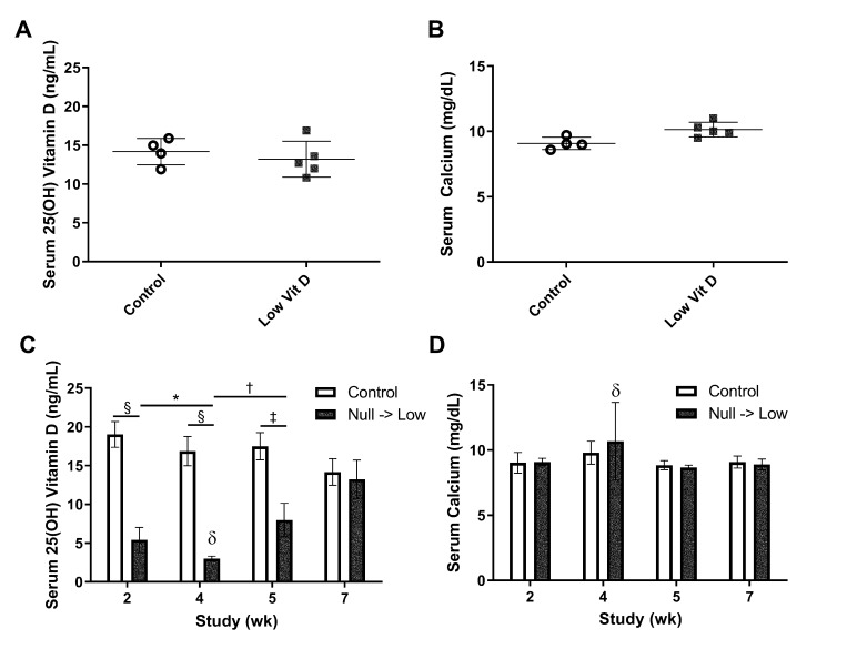
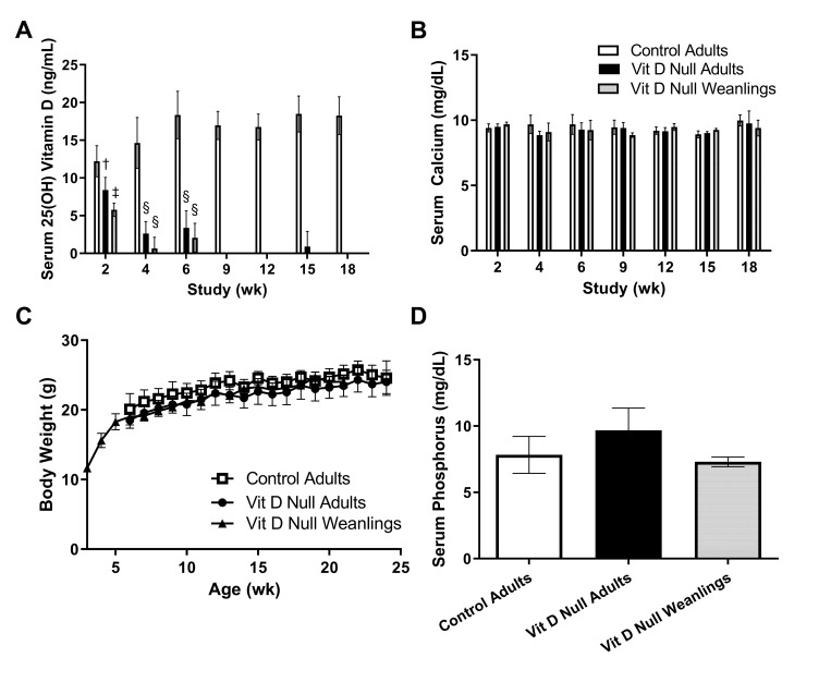
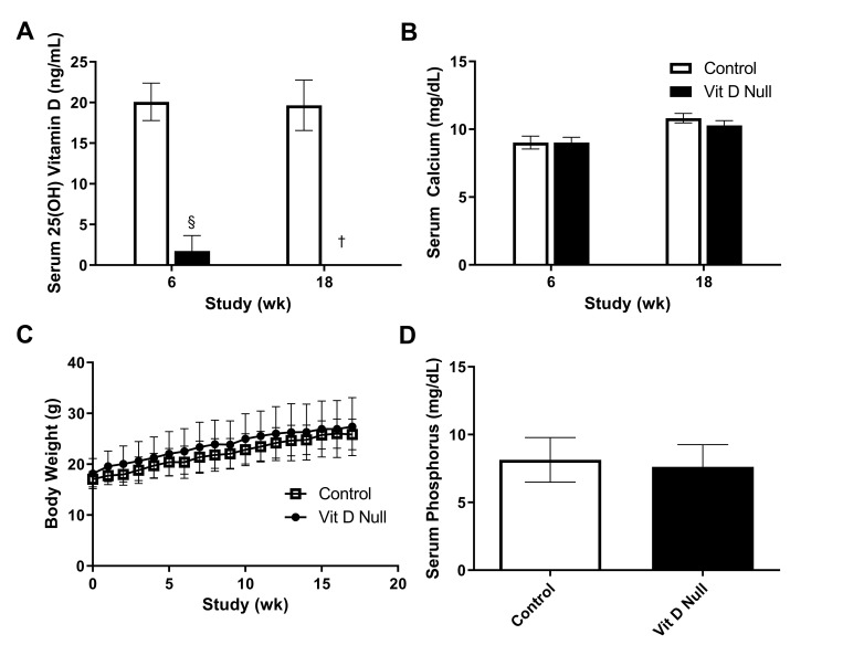
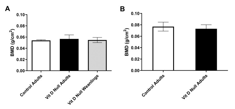
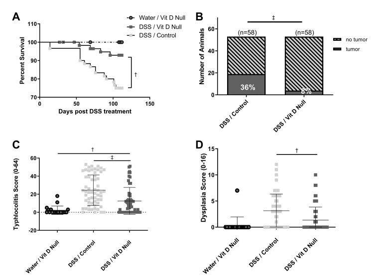
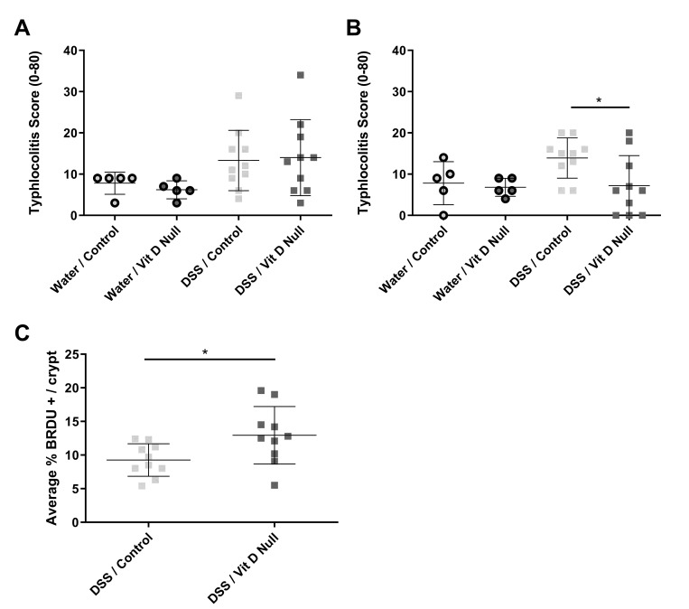
Both human epidemiologic data and animal studies suggest that low serum vitamin D increases the risk of inflammatory bowel disease (IBD) and consequently IBD-associated colorectal cancer. We tested the hypothesis that vitamin D deficiency increases the risk for colitis-associated colon cancer (CAC) by using an established CAC mouse model, 129-Smad3tm1Par/J (Smad3-/-) mice, which have defective transforming growth factor β-signaling and develop colitis and CAC after the administration of dextran sodium sulfate (DSS). After determining a dietary regimen that induced chronic vitamin D deficiency in Smad3-/- mice, we assessed the effects of vitamin D deficiency on CAC. Decreasing dietary vitamin D from 1 IU/g diet (control diet) to 0.2 IU /g diet did not decrease serum 25-hydroxyvitamin D (25(OH)D) levels in Smad3-/- mice. A diet devoid of vitamin D (< 0.02 IU/g diet [no added vitamin D]; vitamin D-null) significantly decreased serum 25(OH)D levels in mice after 2 wk (null compared with control diet: 8.4 mg/mL compared with 12.2 ng/mL) and further decreased serum levels to below the detection limit after 9 wk but did not affect weight gain, serum calcium levels, bone histology, or bone mineral density. In light of these results, Smad3-/- mice were fed a vitamin D-null diet and given DSS to induce colitis. Unexpectedly, DSS-treated Smad3-/- mice fed a vitamin D-null diet had improved survival, decreased colon tumor incidence (8% compared with 36%), and reduced the incidence and severity of colonic dysplasia compared with mice fed the control diet. These effects correlated with increased epithelial cell proliferation and repair early in the disease, perhaps reducing the likelihood of developing chronic colitis and progression to cancer. Our results indicate that vitamin D deficiency is beneficial in some cases of CAC, possibly by promoting intestinal healing.


Figures

Figure 1.
Figure 1.
Low vitamin D diet does not decrease serum vitamin D or serum calcium levels in Smad3−/− mice. Serum 25(OH)D and calcium were evaluated in 2 dietary regimens involving low vitamin D diet. In one regimen, (A) serum 25(OH) D and (B) serum calcium were measured after animals were fed control (n = 4) or vitamin D low (n = 5) diet for 6 wk starting at weaning. In the other regimen, (C) serum 25(OH)D and (D) serum calcium were measured at weeks 2, 4, 5, and 7 from study initiation in mice that received vitamin D-null diet for 4 wk and then were switched to low vitamin D diet compared with those on control diet for the duration of the study. δ indicates the time point at which mice on vitamin D-null diet in the null→low diet treatment group were switched to the low vitamin D diet. Mean and SD are indicated on dot plots. Error bars on bar graphs are SD. *, P < 0.05; †, P < 0.01; ‡, P < 0.001; §, P < 0.0001 (Student's t test) between groups are indicated. Comparisons were made between control compared with null→low group and also between null→low diet groups before and after the diet switch at 4 wk.
Figure 2.
Figure 2.
Vitamin D-null diet significantly decreases serum vitamin D levels without altering serum calcium, weight gain, or serum phosphorus in Smad3−/− mice. (A) Serum 25(OH)D and (B) serum calcium in Smad3−/− mice fed control diet (n = 5, 6-wk-old mice) or vitamin D-null diet (n = 5, 3-wk-old mice [weanling] and n = 5, 6-wk-old mice [adult]). Body weight was measured weekly and (C) average body weight (in grams) is plotted for each group. (D) Serum phosphorus was measured after 18 wk on diet (n = 4 per group). Error bars are SD. One-way ANOVA (parametric) followed by a Bonferroni post hoc test for multiple comparisons. †, P < 0.01; ‡, P < 0.001; §, P < 0.0001 compared with control.
Figure 3.
Figure 3.
Vitamin D-null diet significantly decreases serum vitamin D levels without altering serum calcium, weight gain, or serum phosphorus in wildtype mice. (A) Serum 25(OH)D and (B) serum calcium in wildtype mice fed control (n = 5, 5- to 8-wk-old mice) or vitamin D-null diet (n = 6, 5- to 8-wk-old mice). Body weight was measured weekly, and (C) average body weight (in grams) is plotted for each group. (D) Serum phosphorus was measured at 18 wk after diet initiation. Error bars indicate 1 SD. †, P < 0.01 (Mann–Whitney U test); §, P < 0.0001 (Student t test) compared with control.
Figure 4.
Figure 4.
Vitamin D-null diet does not alter bone density in Smad3−/− or wildtype mice. Total body bone mineral density (BMD) scanning was performed after euthanasia of Smad3−/− and wildtype mice at 18 wk after diet initiation. (A) Smad3−/− mice fed control diet (n = 5, 6-wk-old mice) or vitamin D-null diet (n = 5, 3-wk-old mice [weanling] and n = 5, 6-wk-old mice [adult]) and (B) wildtype mice fed control (n = 5, 5- to 8-wk-old mice) or vitamin D-null diet (n = 6, 5- to 8-wk-old mice). Error bars indicate 1 SD. No significant differences according to Kruskal–Wallis testing.
Figure 5.
Figure 5.
Vitamin D-null diet protects against DSS-induced colitis and colon cancer in Smad3−/− mice. Smad3−/− mice were fed vitamin D-null or control diet and treated with DSS in drinking water (n = 58 mice per group) or with untreated water (n = 20) and followed for disease development until 16 wk after initial DSS treatment, a duration sufficient for CAC development in this model. (A) Survival was compared through log-rank testing. Cecum and colon were assessed histologically at endpoint for evidence of (B) neoplasia, (C) typhlocolitis, and (D) dysplasia. Typhlocolitis and dysplasia scores were compared through Kruskal–Wallis testing followed by Dunn posthoc testing for pairwise comparisons. Means are indicated on dot plots; error bars on bar graphs indicate 1 SD. Dysplasia incidence and neoplasia incidence were compared through Fisher Exact testing. †, P < 0.01; ‡, P < 0.001; §, P < 0.0001.
Figure 6.
Figure 6.
Vitamin D-null diet is associated with increased cell proliferation following DSS-treatment in Smad3−/− mice during the early inflammatory phase of disease. Smad3−/− mice were fed vitamin D-null or control diet and treated with DSS (n = 10 mice per group per time point) or untreated water (n = 5 mice per group per time point). Animals were euthanized at (A) 9 d and (B) 16 d after initiation of DSS treatment, and typhlocolitis scores were generated on the basis of histologic examination of cecal and colonic tissue. Groups were compared by one-way ANOVA (parametric), and pairwise comparisons between groups on the same diet in addition to comparisons between groups in the same treatment (DSS or water) were performed through Bonferroni posthoc testing. (C) BrdU was used to quantify actively proliferating colonic epithelial cells during the healing phase of disease after DSS treatment (16 d after DSS) and compared by using the Mann–Whitney U test. Means (error bars, 1 SD) are indicated on dot plots. *, P < 0.05.

Similar articles

Cited by

References

    1. Aggarwal A, Kallay E. 2016. Cross talk between the calcium-sensing receptor and the vitamin D system in prevention of cancer. Front Physiol 7:1–8. 10.3389/fphys.2016.00451. - DOI - PMC - PubMed
    1. Amling M, Priemel M, Holzmann T, Chapin K, Rueger JM, Baron R, Demay MB. 1999. Rescue of the skeletal phenotype of vitamin D receptor-ablated mice in the setting of normal mineral ion homeostasis: formal histomorphometric and biomechanical analyses. Endocrinology 140:4982–4987. 10.1210/endo.140.11.7110. - DOI - PubMed
    1. Ananthakrishnan AN, Cheng SC, Cai T, Cagan A, Gainer VS, Szolovits P, Shaw SY, Churchill S, Karlson EW, Murphy SN, Kohane I, Liao KP. 2014. Association between reduced plasma 25-hydroxy vitamin D and increased risk of cancer in patients with inflammatory bowel diseases. Clin Gastroenterol Hepatol 12:821–827. 10.1016/j.cgh.2013.10.011. - DOI - PMC - PubMed
    1. Ananthakrishnan AN, Khalili H, Higuchi LM, Bao Y, Korzenik JR, Giovannucci EL, Richter JM, Fuchs CS, Chan AT. 2012. Higher predicted vitamin D status is associated with reduced risk of Crohn's disease. Gastroenterology 142:482–489. 10.1053/j.gastro.2011.11.040. - DOI - PMC - PubMed
    1. Anglin RE, Samaan Z, Walter SD, McDonald SD. 2013. Vitamin D deficiency and depression in adults: systematic review and meta-analysis. Br J Psychiatry 202:100–107. 10.1192/bjp.bp.111.106666. - DOI - PubMed

Publication types