. 2020 Mar 5:101:109645.
doi: 10.1016/j.jbiomech.2020.109645. Epub 2020 Jan 21.

Simulating ventricular systolic motion in a four-chamber heart model with spatially varying robin boundary conditions to model the effect of the pericardium

Affiliations

Simulating ventricular systolic motion in a four-chamber heart model with spatially varying robin boundary conditions to model the effect of the pericardium

Marina Strocchi et al. J Biomech. .

Abstract

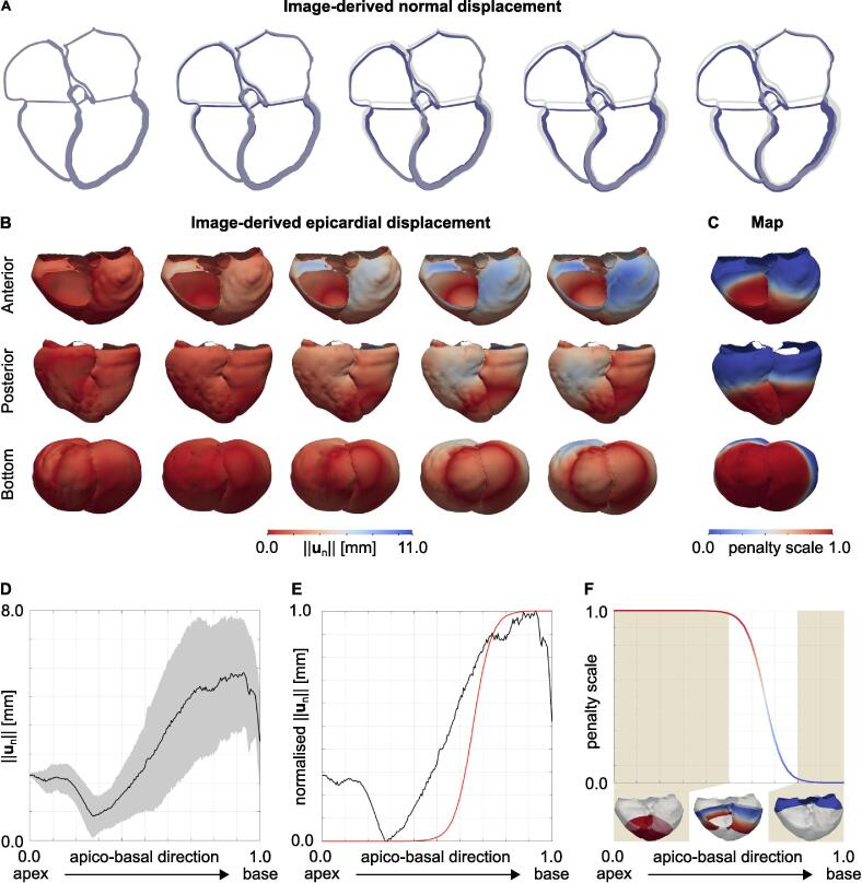
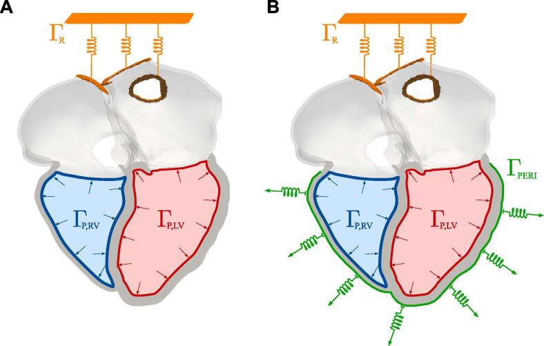
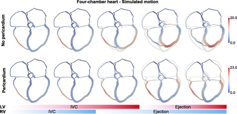
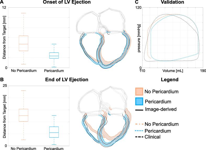
The pericardium affects cardiac motion by limiting epicardial displacement normal to the surface. In computational studies, it is important for the model to replicate realistic motion, as this affects the physiological fidelity of the model. Previous computational studies showed that accounting for the effect of the pericardium allows for a more realistic motion simulation. In this study, we describe the mechanism through which the pericardium causes improved cardiac motion. We simulated electrical activation and contraction of the ventricles on a four-chamber heart in the presence and absence of the effect of the pericardium. We simulated the mechanical constraints imposed by the pericardium by applying normal Robin boundary conditions on the ventricular epicardium. We defined a regional scaling of normal springs stiffness based on image-derived motion from CT images. The presence of the pericardium reduced the error between simulated and image-derived end-systolic configurations from 12.8±4.1 mm to 5.7±2.5 mm. First, the pericardium prevents the ventricles from spherising during isovolumic contraction, reducing the outward motion of the free walls normal to the surface and the upwards motion of the apex. Second, by restricting the inward motion of the free and apical walls of the ventricles the pericardium increases atrioventricular plane displacement by four folds during ejection. Our results provide a mechanistic explanation of the importance of the pericardium in physiological simulations of electromechanical cardiac function.

Keywords: Apico-basal shortening; Cardiac electromechanics; Computer models; Heart failure; Pericardium; Ventricular systolic motion.


Conflict of interest statement

Declaration of Competing Interest The authors have no conflict of interests.

Figures

Fig. 1
Fig. 1
A Target systolic motion. The images represent the motion of a slice of the patient’s heart during systole as computed by the motion tracking algorithm (blue moving geometry). The grey geometry represents the ED geometry. B Epicardial displacement. The images show the distribution of the displacement of the epicardium normal to the surface during systole in an anterior (top row), posterior (middle row) and bottom (bottom row) view. C Penalty map. The figure shows the anterior, posterior and bottom view of the penalty map for the displacement normal to the surface applied on the epicardium of the ventricles to model the effect of the pericardium on the ventricles. D Apex to base epicardial displacement. The plot shows the epicardial displacement normal to the surface against the apico-basal direction (0 at the base and 1 at the apex). The black line shows the average trend, while the gray area shows the standard deviation. E Apex to base normalised epicardial displacement. The plot shows the epicardial displacement normal to the surface normalised between 0 and 1, together with the function we used to define the scale for the spring stiffness (red line). F Apex to base penalty map. The plot shows the penalty scale we derived from the data against the apico-basal direction. The function was computed by flipping the red curve shown in panel E. Epicardial regions with low and high displacement normal to the surface were applied with maximum and minimum penalty, respectively.
Fig. 2
Fig. 2
Boundary conditions. Boundary conditions applied in the simulation without A and with B the effect of the pericardium. We applied omni-directional springs at the two cropped right pulmonary veins and at the cropped superior vena cava, represented by the orange boundary ΓR. Neumann boundary conditions for left and right ventricular pressure were applied at the left ventricular endocardium ΓP,LV (red) and at the right ventricular endocardium ΓP,RV (blue), respectively. In the simulation with the pericardium, we added normal springs on the epicardial surface of the ventricles ΓPERI, shown in green. (For interpretation of the references to colour in this figure legend, the reader is referred to the web version of this article.)
Fig. 3
Fig. 3
Simulated motion. The images show the motion of a slice of the four-chamber geometry for the simulation without (top row) and with (bottom row) the pericardium. The grey geometry represents the ED configuration, while the moving geometry is coloured according to the displacement magnitude. The blue and red bars at the bottom display the phase of the cardiac cycle for the RV and the LV, respectively. (For interpretation of the references to colour in this figure legend, the reader is referred to the web version of this article.)
Fig. 4
Fig. 4
A, B Comparison with image-derived displacement. We show the comparison between the image-derived and the simulated motion. Figure A and B show the comparison at the onset of LV ejection and at the end of LV ventricular systole. For each figure, On the left we show the boxplots of the Euclidean distance between the image-derived and the simulated motion without (orange) and with (light-blue) the effect of the pericardium. On the right, we overlapped the image-derived configuration (geometry with black borders) to the configurations simulated without (orange) and with (light-blue) the effect of the pericardium. C Pressure-volue loop validation. The clinical pressure–volume loop (black dashed line) is compared to the simulated pressure–volume loops without (orange) and with (light-blue) the effect of the pericardium. (For interpretation of the references to colour in this figure legend, the reader is referred to the web version of this article.)

Similar articles

Cited by

References

    1. Abbasi A.S., Eber L.M., Macalpin R.N., Kattus A.A. Paradoxical motion of interventricular septum in left bundle branch block. Circulation. 1974;49:423–427. - PubMed
    1. Aguado-Sierra J., Krishnamurthy A., Villongco C., Chuang J., Howard E., Gonzales M.J., Omens J., Krummen D.E., Narayan S., Kerckhoffs R.C. Patient-specific modeling of dyssynchronous heart failure: a case study. Progress Biophys. Molecul. Biol. 2011;107:147–155. - PMC - PubMed
    1. Augustin C.M., Crozier A., Neic A., Prassl A.J., Karabelas E., Ferreira da Silva T., Fernandes J.F., Campos F., Kuehne T., Plank G. Patient-specific modeling of left ventricular electromechanics as a driver for haemodynamic analysis. EP Europace. 2016;18:iv121–iv129. - PMC - PubMed
    1. Augustin C.M., Neic A., Liebmann M., Prassl A.J., Niederer S.A., Haase G., Plank G. Anatomically accurate high resolution modeling of human whole heart electromechanics: a strongly scalable algebraic multigrid solver method for nonlinear deformation. J. Comput. Phys. 2016;305:622–646. - PMC - PubMed
    1. Baillargeon B., Rebelo N., Fox D.D., Taylor R.L., Kuhl E. The living heart project: a robust and integrative simulator for human heart function. Eur. J. Mech. A Solids. 2014;48:38–47. - PMC - PubMed

Publication types