

Effects of repetitive transcranial magnetic stimulation on resting-state connectivity: A systematic review
- PMID: 32014552
- PMCID: PMC7571509
- DOI: 10.1016/j.neuroimage.2020.116596
Effects of repetitive transcranial magnetic stimulation on resting-state connectivity: A systematic review
Abstract
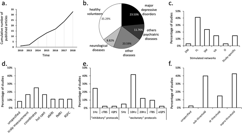
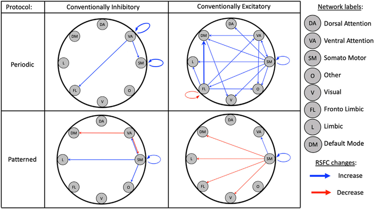
The brain is organized into networks that reorganize dynamically in response to cognitive demands and exogenous stimuli. In recent years, repetitive transcranial magnetic stimulation (rTMS) has gained increasing use as a noninvasive means to modulate cortical physiology, with effects both proximal to the stimulation site and in distal areas that are intrinsically connected to the proximal target. In light of these network-level neuromodulatory effects, there has been a rapid growth in studies attempting to leverage information about network connectivity to improve neuromodulatory control and intervention outcomes. However, the mechanisms-of-action of rTMS on network-level effects remain poorly understood and is based primarily on heuristics from proximal stimulation findings. To help bridge this gap, the current paper presents a systematic review of 33 rTMS studies with baseline and post-rTMS measures of fMRI resting-state functional connectivity (RSFC). Literature synthesis revealed variability across studies in stimulation parameters, studied populations, and connectivity analysis methodology. Despite this variability, it is observed that active rTMS induces significant changes on RSFC, but the prevalent low-frequency-inhibition/high-frequency-facilitation heuristic endorsed for proximal rTMS effects does not fully describe distal connectivity findings. This review also points towards other important considerations, including that the majority of rTMS-induced changes were found outside the stimulated functional network, suggesting that rTMS effects tend to spread across networks. Future studies may therefore wish to adopt conventions and systematic frameworks, such as the Yeo functional connectivity parcellation atlas adopted here, to better characterize network-level effect that contribute to the efficacy of these rapidly developing noninvasive interventions.
Keywords: Distal effects; Network neuroscience; Repetitive transcranial magnetic stimulation; Resting-state functional connectivity.
Copyright © 2020 The Authors. Published by Elsevier Inc. All rights reserved.
Conflict of interest statement
Declaration of competing interest The authors do not have any conflict of interest to declare.
Figures
References
-
- Baeken C, Duprat R, Wu GR, De Raedt R, van Heeringen K, 2017. Subgenual anterior cingulate-medial orbitofrontal functional connectivity in medication-resistant major depression: a neurobiological marker for accelerated intermittent theta burst stimulation treatment? Biol Psychiatry Cogn Neurosci Neuroimaging 2 (7), 556–565. 10.1016/j.bpsc.2017.01.001. - DOI - PubMed
-
- Baeken C, Marinazzo D, Wu G-R, Van Schuerbeek P, De Mey J, Marchetti I, De Raedt R, 2014. Accelerated HF-rTMS in treatment-resistant unipolar depression: insights from subgenual anterior cingulate functional connectivity. World J. Biol. Psychiatr 15 (4), 286–297. - PubMed
Publication types
MeSH terms
Grants and funding
LinkOut - more resources
Full Text Sources
Medical
Miscellaneous
