. 2020 Mar 23;88(4):e00886-19.
doi: 10.1128/IAI.00886-19. Print 2020 Mar 23.

Activation of ASC Inflammasome Driven by Toll-Like Receptor 4 Contributes to Host Immunity against Rickettsial Infection

Affiliations

Activation of ASC Inflammasome Driven by Toll-Like Receptor 4 Contributes to Host Immunity against Rickettsial Infection

Claire Rumfield et al. Infect Immun. .

Abstract

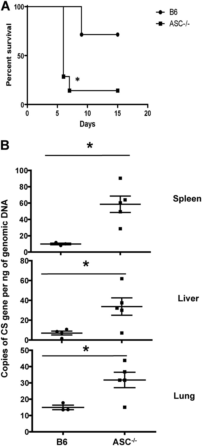
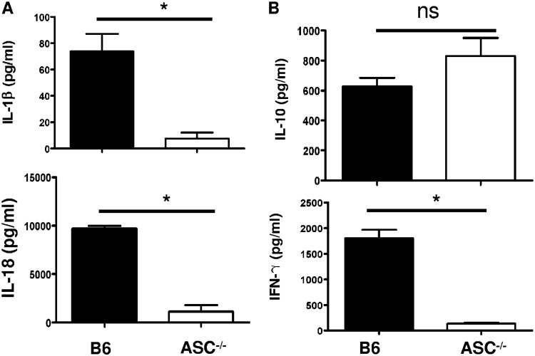
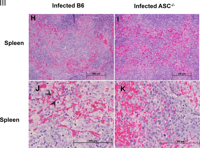
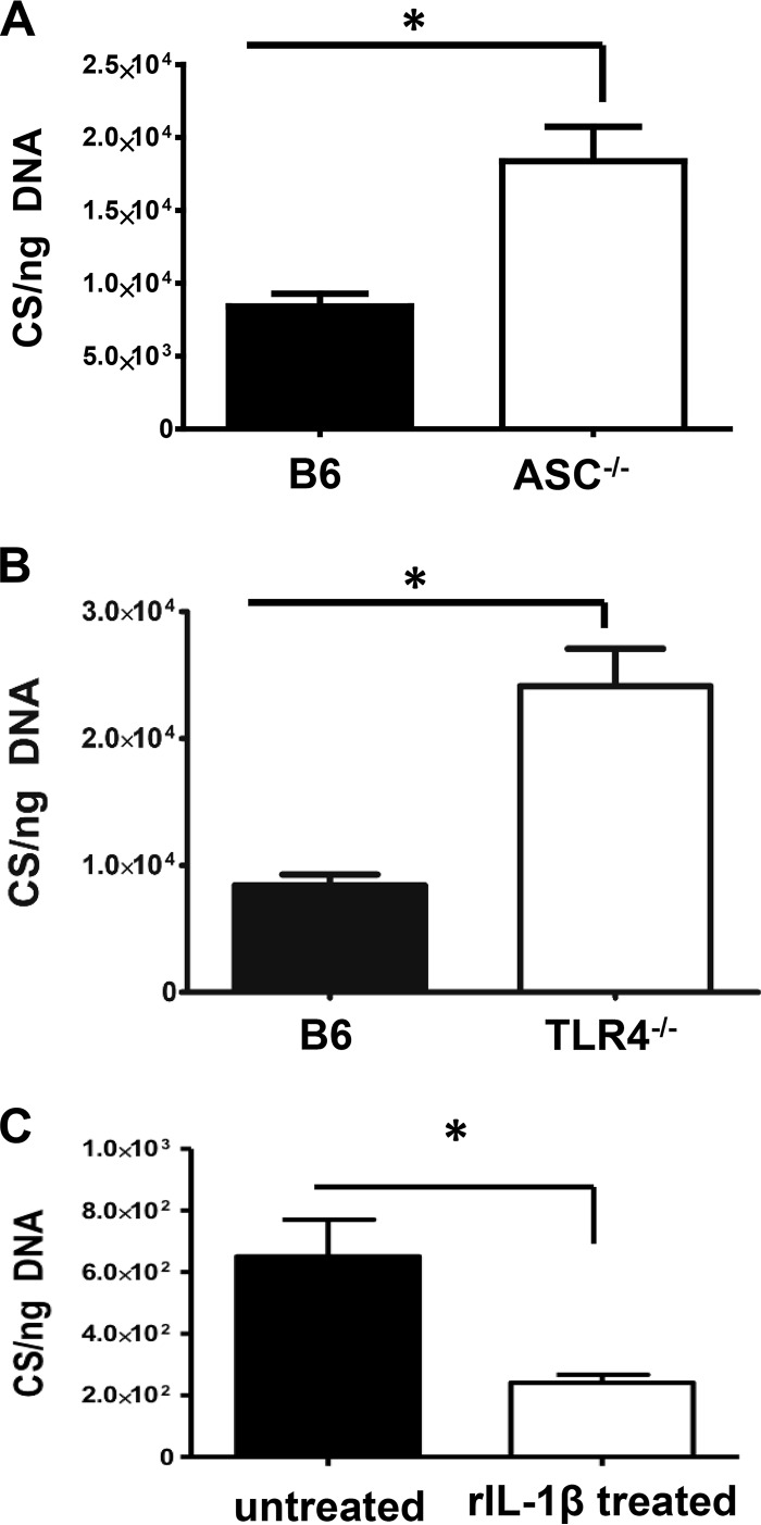
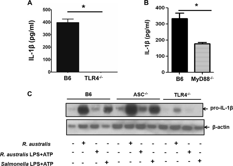
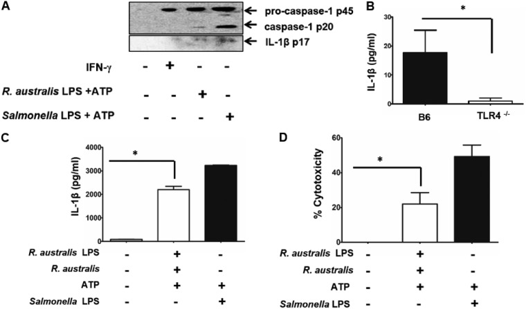
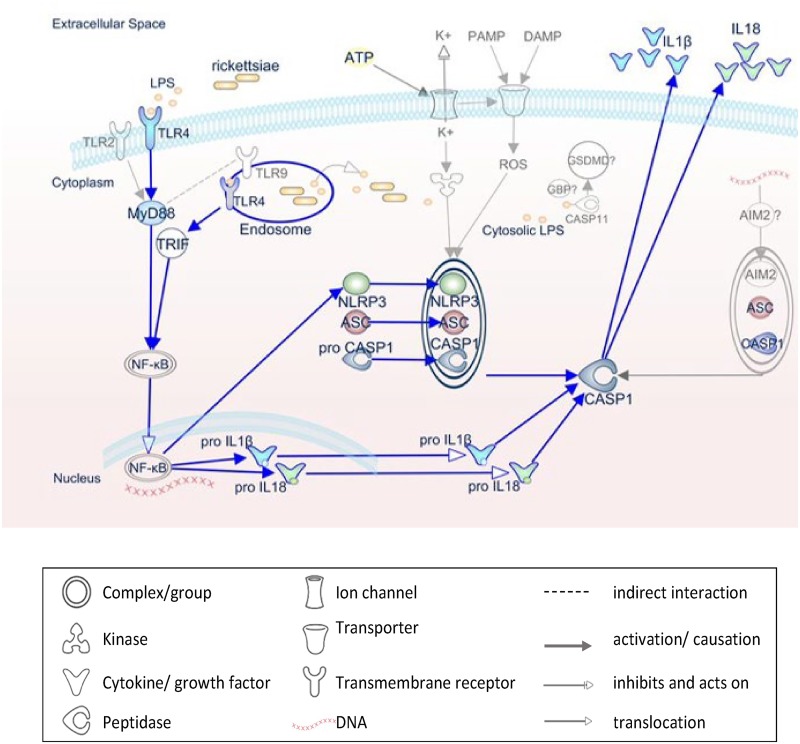
Rickettsiae are cytosolically replicating, obligately intracellular bacteria causing human infections worldwide with potentially fatal outcomes. We previously showed that Rickettsia australis activates ASC inflammasome in macrophages. In the present study, host susceptibility of ASC inflammasome-deficient mice to R. australis was significantly greater than that of C57BL/6 (B6) controls and was accompanied by increased rickettsial loads in various organs. Impaired host control of R. australis in vivo in ASC-/- mice was associated with dramatically reduced levels of interleukin 1β (IL-1β), IL-18, and gamma interferon (IFN-γ) in sera. The intracellular concentrations of R. australis in bone marrow-derived macrophages (BMMs) of TLR4-/- and ASC-/- mice were significantly greater than those in BMMs of B6 controls, highlighting the important role of inflammasome and these molecules in controlling rickettsiae in macrophages. Compared to B6 BMMs, TLR4-/- BMMs failed to secrete a significant level of IL-1β and had reduced expression levels of pro-IL-1β in response to infection with R. australis, suggesting that rickettsiae activate ASC inflammasome via a Toll-like receptor 4 (TLR4)-dependent mechanism. Further mechanistic studies suggest that the lipopolysaccharide (LPS) purified from R. australis together with ATP stimulation led to cleavage of pro-caspase-1 and pro-IL-1β, resulting in TLR4-dependent secretion of IL-1β. Taken together, these observations indicate that activation of ASC inflammasome, most likely driven by interaction of TLR4 with rickettsial LPS, contributes to host protective immunity against R. australis These findings provide key insights into defining the interactions of rickettsiae with the host innate immune system.

Keywords: ASC; LPS; TLR4; inflammasome; rickettsiae.


Figures

FIG 1
FIG 1
ASC inflammasome contributes significantly to host susceptibility to and in vivo clearance of R. australis. (A) B6 and ASC−/− mice were inoculated with 0.5 LD50 of R. australis i.v. Host survival was monitored daily until day 20 p.i. Each group of mice included 6 to 10 mice. (B) On day 4 p.i., mice were sacrificed, and the concentrations of rickettsiae in spleens, livers, and lungs were quantified by real-time PCR. Data are means ± standard errors (SE) for 5 mice for each group. Data represent two independent experiments. *, P < 0.05 for a significant difference between B6 and ASC−/− mice.
FIG 2
FIG 2
ASC inflammasome is indispensable for significant serum levels of inflammasome-mediated cytokines during rickettsial infection. B6 and ASC−/− mice were inoculated with 0.5 LD50 of R. australis i.v. On day 4 p.i., mice were sacrificed. (A) The in vivo production of inflammasome-mediated cytokines, including IL-1β and IL-18, was assessed in sera by ELISA. (B) Serum levels of IL-10 and IFN-γ were measured by ELISA. Results are means ± SE of data from 4 to 8 mice per group. Data represent two independent experiments. ns, not significant. *, P < 0.05 for a significant difference between B6 and ASC−/− mice.
FIG 3
FIG 3
Histopathological analysis of tissues of R. australis-infected B6 and ASC−/− mice. B6 and ASC−/− mice were inoculated with 0.5 LD50 of R. australis i.v. On day 4 p.i., mice were euthanized, and tissues were collected. (I) As negative controls, uninfected B6 and ASC−/− mice were monitored and euthanized along with the infected mouse groups. Histopathological analysis of livers and spleens was performed at magnifications of ×10 (scale bar, 200 μm) and ×40 (scale bar, 100 μm). (II) (A and B) Foci of inflammatory cells in liver tissue (arrows). (C to E) Perivascular infiltration (arrows) and polymorphonuclear neutrophils (PMNs) (arrowheads) in livers. (F and G) Coagulative necrotic infarct (arrowheads) only in livers of infected ASC−/− mice and surrounding inflammation and tissue repair (arrows). (III) (H to K) Spleens of infected B6 and ASC−/− mice. Clusters of macrophages and normal periarteriolar lymphocyte sheaths, a portion of white pulp, in spleens of both groups of mice are shown. In red pulp, PMNs (arrowheads) were found only in infected B6 mice (H and J), not in infected ASC−/− mice (I and K).
FIG 3
FIG 3
Histopathological analysis of tissues of R. australis-infected B6 and ASC−/− mice. B6 and ASC−/− mice were inoculated with 0.5 LD50 of R. australis i.v. On day 4 p.i., mice were euthanized, and tissues were collected. (I) As negative controls, uninfected B6 and ASC−/− mice were monitored and euthanized along with the infected mouse groups. Histopathological analysis of livers and spleens was performed at magnifications of ×10 (scale bar, 200 μm) and ×40 (scale bar, 100 μm). (II) (A and B) Foci of inflammatory cells in liver tissue (arrows). (C to E) Perivascular infiltration (arrows) and polymorphonuclear neutrophils (PMNs) (arrowheads) in livers. (F and G) Coagulative necrotic infarct (arrowheads) only in livers of infected ASC−/− mice and surrounding inflammation and tissue repair (arrows). (III) (H to K) Spleens of infected B6 and ASC−/− mice. Clusters of macrophages and normal periarteriolar lymphocyte sheaths, a portion of white pulp, in spleens of both groups of mice are shown. In red pulp, PMNs (arrowheads) were found only in infected B6 mice (H and J), not in infected ASC−/− mice (I and K).
FIG 4
FIG 4
Contributions of inflammasome components to host control of intracellular R. australis in macrophages. BMMs of B6, ASC−/− (A), and TLR4−/− (B) mice were infected with R. australis at an MOI of 2. Fresh medium was replaced every 24 h after infection. (C) RAW 264.7 macrophages were cultured and treated with 10 ng/ml recombinant IL-1β at the time of infection. At 48 h p.i., cells were washed, and the total DNA was extracted. The concentrations of rickettsiae in these mouse macrophages were evaluated by quantitative real-time PCR at 48 h p.i. The number of citrate synthase (CS) gene copies per ng of genomic DNA represents the quantity of rickettsiae. Data represent two independent experiments. *, P < 0.05.
FIG 5
FIG 5
Mechanisms involved in mediating the secretion of IL-1β by R. australis-infected macrophages. BMMs were isolated from B6, TLR4−/−, MyD88−/−, and ASC−/− mice. BMMs were infected with R. australis at an MOI of 6 or stimulated with 50 ng/ml of purified R. australis LPS plus ATP for 24 h. ATP was added 1 h prior to collection. Supernatant was collected from infected BMMs of B6, TLR4−/− (A), and MyD88−/− (B) mice. Levels of secretion of IL-1β by these infected macrophages were evaluated by ELISA. (C) The synthesis of pro-IL-1β in BMMs of B6, ASC−/− and TLR4−/− mice was evaluated by specific antibodies against pro-IL-1β by immunoblotting after the cell lysates were collected. Salmonella LPS plus ATP served as the positive control. Data represent two independent experiments.
FIG 6
FIG 6
Rickettsial LPS serves as an efficient stimulus for activating inflammasome in macrophages. Rickettsial LPS was isolated and purified. BMMs were isolated from B6 and TLR4−/− mice. Rickettsial LPS was added to BMMs at a concentration of 100 ng/ml for 24 h. ATP (5 mM) was added to the cell culture for 1 h prior to collection. Salmonella LPS (100 ng/ml) plus ATP served as the positive control. (A) Supernatants of B6 BMMs stimulated with rickettsial LPS and ATP were collected and then concentrated. Activation of inflammasome was evaluated by immunoblotting using specific antibodies against pro-caspase-1 (p45), activated caspase-1 (p20), and activated IL-1β (p17). BMMs stimulated with IFN-γ (40 ng/ml) served as controls. (B) Supernatants of BMMs from B6 and TLR4−/− mice stimulated with purified LPS of R. australis and ATP were collected. Secretion levels of IL-1β by these stimulated cells were measured by ELISA. Next, B6 BMMs were first primed with 100 ng/ml rickettsial LPS and then infected with R. australis at an MOI of 6. Then, 5 mM ATP was added to the cells for 1 h prior to collection of the supernatant. Secretion levels of IL-1β was determined by ELISA (C). The cytotoxicity was determined by LDH assay (D). Data represent two independent experiments. *, P < 0.05.
FIG 7
FIG 7
Schematic diagram of inflammasome activation by rickettsiae. This IPA-generated schematic depicts the molecular mechanisms involved in the activation of inflammasome by pathogen-associated molecular patterns during rickettsial infection. R. australis is recognized by and triggers the host’s NLRP3/ASC inflammasome, which leads to caspase-1 activation and secretion of IL-1β and IL-18 and culminates in the control of bacterial replication in macrophages and in vivo. In murine macrophages, whereas NLRP3 is dispensable for inflammasome activation by R. australis, AIM2/ASC is hypothesized to play a role. The priming signal for NLRP3 inflammasome activation is provided by recognition of rickettsial LPS by host TLR4 in both MyD88-dependent and -independent mechanisms. Rickettsiae invade the host cytosol to provide signal 2 for NLRP3/ASC inflammasome through an unknown molecule. Pathways demonstrated in our studies are blue, while mechanisms hypothesized but not tested are gray.

References

    1. Binder AM, Nichols Heitman K, Drexler NA. 2019. Diagnostic methods used to classify confirmed and probable cases of spotted fever rickettsioses—United States, 2010–2015. MMWR Morb Mortal Wkly Rep 68:243–246. doi:10.15585/mmwr.mm6810a3. - DOI - PMC - PubMed
    1. Drexler NA, Yaglom H, Casal M, Fierro M, Kriner P, Murphy B, Kjemtrup A, Paddock CD. 2017. Fatal Rocky Mountain spotted fever along the United States-Mexico Border, 2013–2016. Emerg Infect Dis 23:1621–1626. doi:10.3201/eid2310.170309. - DOI - PMC - PubMed
    1. Takeuchi O, Akira S. 2010. Pattern recognition receptors and inflammation. Cell 140:805–820. doi:10.1016/j.cell.2010.01.022. - DOI - PubMed
    1. Guillotte ML, Gillespie JJ, Chandler CE, Rahman MS, Ernst RK, Azad AF. 2018. Rickettsia lipid A biosynthesis utilizes the late acyltransferase LpxJ for secondary fatty acid addition. J Bacteriol 200:e00334-18. doi:10.1128/JB.00334-18. - DOI - PMC - PubMed
    1. Smalley C, Bechelli J, Rockx-Brouwer D, Saito T, Azar SR, Ismail N, Walker DH, Fang R. 2016. Rickettsia australis activates inflammasome in human and murine macrophages. PLoS One 11:e0157231. doi:10.1371/journal.pone.0157231. - DOI - PMC - PubMed

Publication types

Supplementary concepts

LinkOut - more resources