. 2020 Mar 23;88(4):e00353-19.
doi: 10.1128/IAI.00353-19. Print 2020 Mar 23.

Atypical Salmonella enterica Serovars in Murine and Human Macrophage Infection Models

Affiliations

Atypical Salmonella enterica Serovars in Murine and Human Macrophage Infection Models

Daniel Hurley et al. Infect Immun. .

Abstract

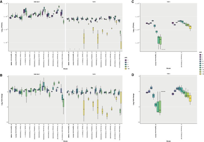
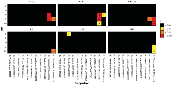
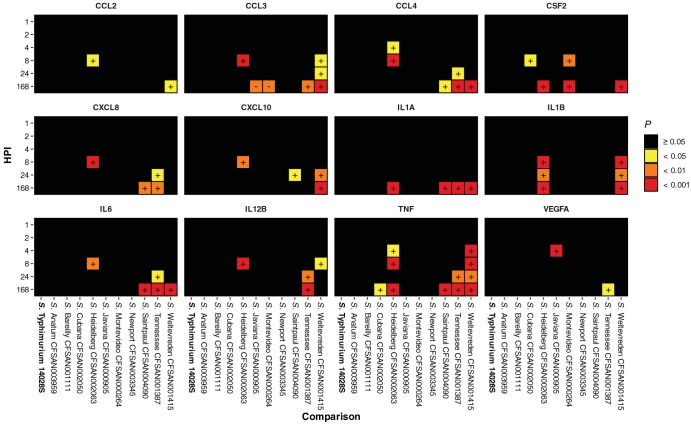
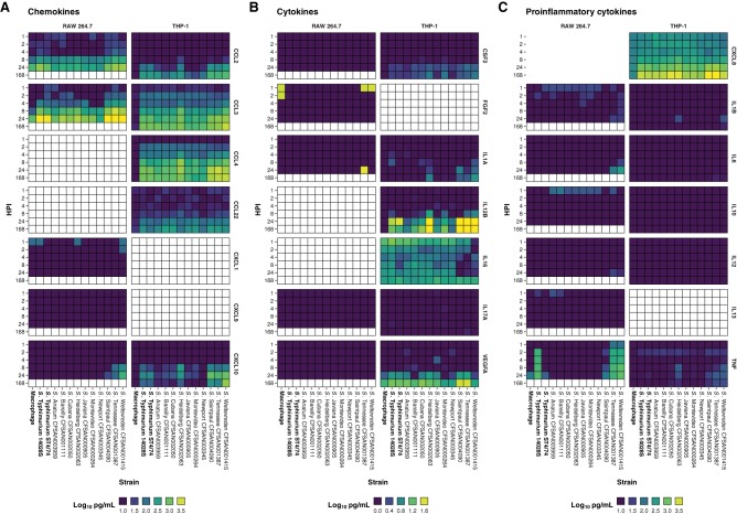
Nontyphoidal Salmonella species are globally disseminated pathogens and are the predominant cause of gastroenteritis. The pathogenesis of salmonellosis has been extensively studied using in vivo murine models and cell lines, typically challenged with Salmonella enterica serovar Typhimurium. Although S. enterica serovars Enteritidis and Typhimurium are responsible for most of the human infections reported to the Centers for Disease Control and Prevention (CDC), several other serovars also contribute to clinical cases of salmonellosis. Despite their epidemiological importance, little is known about their infection phenotypes. Here, we report the virulence characteristics and genomes of 10 atypical S. enterica serovars linked to multistate foodborne outbreaks in the United States. We show that the murine RAW 264.7 macrophage model of infection is unsuitable for inferring human-relevant differences in nontyphoidal Salmonella infections, whereas differentiated human THP-1 macrophages allowed these isolates to be further characterized in a more human-relevant context.

Keywords: Salmonella; Salmonella enterica; cytokines; infection; macrophages; pathogenicity; virulence; whole-genome sequencing.


Figures

FIG 1
FIG 1
Survival of isolates following phagocytosis by RAW 264.7 and THP-1 macrophages, assessed by gentamicin protection assay. (A) Survival of isolates at 2, 4, 8, and 24 hours postinfection (hpi) following phagocytosis by RAW 264.7 macrophages and at 2, 4, 8, 24, and 168 hpi following phagocytosis by THP-1 macrophages, reported as log10 CFU/ml. (B) Survival of isolates at 4, 8, and 24 hpi following phagocytosis by RAW 264.7 macrophages and at 4, 8, 24, and 168 hpi following phagocytosis by THP-1 macrophages, reported as log2 fold change compared to survival at 2 hpi. Results for isolates in the study correspond to the mean of three independent assays (n = 3) with duplicate technical replicates. Results for reference Salmonella enterica serovar Typhimurium strains 14028S and ST4/74 correspond to the mean of six independent assays (n = 6) with duplicate technical replicates. (C) Survival at 2, 4, 8, 24, 48, 72, 96, 120, 144, and 168 hpi of S. enterica Tennessee CFSAN001387, in comparison with S. Typhimurium ST4/74, following phagocytosis by THP-1 macrophages, reported as log10 CFU/ml. (D) Survival at 4, 8, 24, 48, 72, 96, 120, 144, and 168 hpi of S. Tennessee CFSAN001387, in comparison with S. Typhimurium ST4/74, following phagocytosis by THP-1 macrophages, reported as log2 fold change compared to survival at 2 hpi. Results correspond to the mean of three independent assays (n = 3) with triplicate technical replicates. The lower and upper hinges correspond to the 25th and 75th percentiles, with the whiskers extending ±1.5 times the range between first and third quartiles.
FIG 2
FIG 2
Significant RAW 264.7 chemokine and proinflammatory cytokine release following infection with isolates compared to that following S. Typhimurium ST4/74 infection. Increased release of chemokine and proinflammatory cytokine proteins by RAW 264.7 macrophages at 1, 2, 4, 8, and 24 hpi with isolates compared to that following infection with S. Typhimurium ST4/74, as determined by one-way analysis of variance (ANOVA). Values correspond to adjusted probability (P) as determined by post hoc analysis of significance using Tukey’s range test. Differences were deemed significant by arbitrary cutoffs at P < 0.05, 0.01, and 0.001. Quantification was determined using a multiplex magnetic bead-based enzyme-linked immunosorbent assay (ELISA) and the Luminex 200 xMAP platform (see Table S7 in the supplemental material).
FIG 3
FIG 3
Significant THP-1 chemokine, cytokine, and proinflammatory cytokine release following infection with isolates compared to that following S. Typhimurium ST4/74 infection. Increased release of chemokine, cytokine, and proinflammatory cytokine proteins by THP-1 macrophages at 1, 2, 4, 8, 24, and 168 hpi with isolates compared to that following infection with S. Typhimurium ST4/74, as determined by one-way ANOVA. Values correspond to adjusted probability (P) as determined by post hoc analysis of significance using Tukey’s range test. Differences were deemed significant by arbitrary cutoffs at P < 0.05, 0.01, and 0.001. Quantification was determined using an electrochemiluminescence-based ELISA method and the Sector Imager 2400 platform (Meso Scale Discovery) (Table S7).
FIG 4
FIG 4
Chemokine, cytokine, and proinflammatory cytokine release from infected RAW 264.7 and THP-1 macrophages. Release of chemokine (A), cytokine (B), and proinflammatory cytokine (C) targets by RAW 264.7 and THP-1 macrophages following infection with isolates, reported as log10 pg/ml. Cells colored white correspond to time points not assayed with respect to protein or cell line. Quantification was determined as described in the “Cytokine quantification” section of Materials and Methods (Table S7).
FIG 5
FIG 5
Sequence variability within SPI-1 to SPI-5. The amino acid sequence similarity for the SPI gene content of each isolate included in this study was compared with that of S. Typhimurium ST4/74.

References

    1. Kirk MD, Pires SM, Black RE, Caipo M, Crump JA, Devleesschauwer B, Döpfer D, Fazil A, Fischer-Walker CL, Hald T, Hall AJ, Keddy KH, Lake RJ, Lanata CF, Torgerson PR, Havelaar AH, Angulo FJ. 2015. World Health Organization estimates of the global and regional disease burden of 22 foodborne bacterial, protozoal, and viral diseases, 2010: a data synthesis. PLoS Med 12:e1001921. doi:10.1371/journal.pmed.1001921. - DOI - PMC - PubMed
    1. Crump JA, Luby SP, Mintz ED. 2004. The global burden of typhoid fever. Bull World Health Organ 82:346–353. - PMC - PubMed
    1. Majowicz SE, Musto J, Scallan E, Angulo FJ, Kirk M, O’Brien SJ, Jones TF, Fazil A, Hoekstra RM, International Collaboration on Enteric Disease “Burden of Illness” Studies. 2010. The global burden of nontyphoidal Salmonella gastroenteritis. Clin Infect Dis 50:882–889. doi:10.1086/650733. - DOI - PubMed
    1. Acheson D, Hohmann EL. 2001. Nontyphoidal salmonellosis. Clin Infect Dis 32:263–269. doi:10.1086/318457. - DOI - PubMed
    1. Ao TT, Feasey NA, Gordon MA, Keddy KH, Angulo FJ, Crump JA. 2015. Global burden of invasive nontyphoidal Salmonella disease, 2010. Emerg Infect Dis 21:941–949. doi:10.3201/eid2106.140999. - DOI - PMC - PubMed

Publication types

LinkOut - more resources