. 2020 Mar 20;295(12):3783-3793.
doi: 10.1074/jbc.RA119.012224. Epub 2020 Feb 3.

A thermophilic phage uses a small terminase protein with a fixed helix-turn-helix geometry

Affiliations

A thermophilic phage uses a small terminase protein with a fixed helix-turn-helix geometry

Janelle A Hayes et al. J Biol Chem. .

Abstract

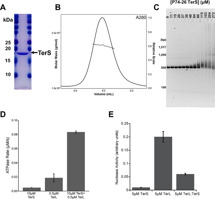
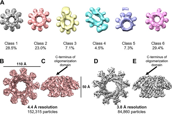
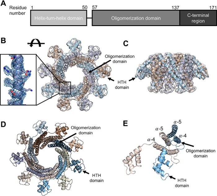
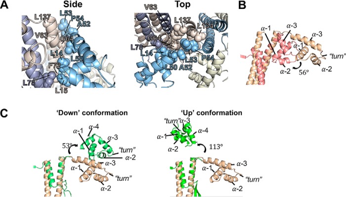
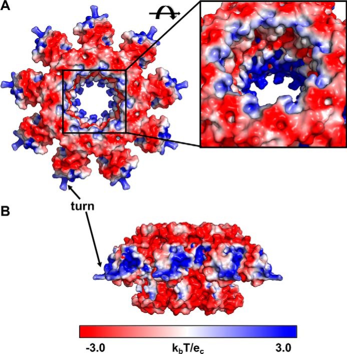
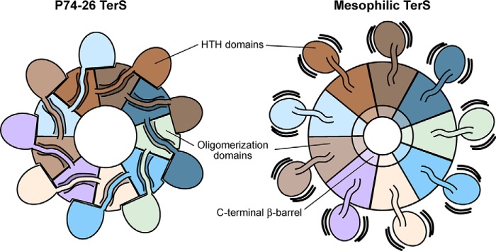
Tailed bacteriophages use a DNA-packaging motor to encapsulate their genome during viral particle assembly. The small terminase (TerS) component of this DNA-packaging machinery acts as a molecular matchmaker that recognizes both the viral genome and the main motor component, the large terminase (TerL). However, how TerS binds DNA and the TerL protein remains unclear. Here we identified gp83 of the thermophilic bacteriophage P74-26 as the TerS protein. We found that TerSP76-26 oligomerizes into a nonamer that binds DNA, stimulates TerL ATPase activity, and inhibits TerL nuclease activity. A cryo-EM structure of TerSP76-26 revealed that it forms a ring with a wide central pore and radially arrayed helix-turn-helix domains. The structure further showed that these helix-turn-helix domains, which are thought to bind DNA by wrapping the double helix around the ring, are rigidly held in an orientation distinct from that seen in other TerS proteins. This rigid arrangement of the putative DNA-binding domain imposed strong constraints on how TerSP76-26 can bind DNA. Finally, the TerSP76-26 structure lacked the conserved C-terminal β-barrel domain used by other TerS proteins for binding TerL. This suggests that a well-ordered C-terminal β-barrel domain is not required for TerSP76-26 to carry out its matchmaking function. Our work highlights a thermophilic system for studying the role of small terminase proteins in viral maturation and presents the structure of TerSP76-26, revealing key differences between this thermophilic phage and its mesophilic counterparts.

Keywords: DNA packaging; DNA recognition; DNA-binding protein; bacteriophage; cryo-EM; helix–turn–helix domain; molecular motor; small terminase; thermophile; viral motor.


Conflict of interest statement

The authors declare that they have no conflicts of interest with the contents of this article

Figures

Figure 1.
Figure 1.
Characterization of TerS gp83. A, SDS-PAGE gel of purified P74-26 gp83. B, SEC-MALS of P74-26 gp83. The UV absorbance at 280-nm wavelength is shown. The measured molecular mass of the complex is 170 kDa, compared with 171 kDa calculated from sequence of a 9-mer. The polydispersity index is 1.000. C, P74-26 gp83 binds DNA with weak affinity. Titrating P74-26 gp83 from 0 to 272 μm (monomer) with 50 ng of the P74-26 gp83 gene shows that TerS has a low affinity for DNA. D, P74-26 gp83 increases the ATPase activity of TerLP74-26 4.4-fold (n = 3, error bars indicate the standard deviation of replicates). E, P74-26 gp83 decreases TerLP74-26 nuclease activity 3.3-fold (n = 3, error bars indicate the standard deviation of replicates).
Figure 2.
Figure 2.
3D Cryo-EM reconstruction of TerSP74-26. A, asymmetric 3D classification shows 9-fold symmetry in the TerSP74-26 ring. B, 4.4 Å resolution asymmetric 3D reconstruction of the TerSP74-26 ring (top). C, side view of asymmetric TerS reconstruction. D, 3.8 Å resolution C9 symmetric 3D reconstruction of the TerSP74-26 ring (top). E, side view of symmetric TerS reconstruction.
Figure 3.
Figure 3.
Model of TerSP74-26. A, TerSP74-26 is comprised of an N-terminal helix–turn–helix domain, a central oligomerization domain, and a C-terminal region. B, built atomic model in 3.8 Å resolution TerSP74-26 symmetric reconstruction (top). Inset, model built into the density of the oligomerization domain. C, side view of the atomic model in the TerSP74-26 reconstruction. D, top view of the atomic model, with the HTH and oligomerization domains indicated. E, in each subunit, α-helix 5 packs into the crevice formed by α-helices 4 and 5 in the counterclockwise subunit. For simplicity, only two subunits (tan and light blue) are shown.
Figure 4.
Figure 4.
The TerSP74-26 linker plays an important role in subunit oligomerization and positions the HTH domain differently than mesophilic TerS proteins. A, hydrophobic residues (labeled) line the linker (residues 51–56) and HTH–oligomerization interfaces between subunits, forming a strong hydrophobic core. B, alignment of the symmetric TerSP74-26 model (tan) with TerSSf6 (pink, PDB code 3HEF) shows that the TerSSf6 HTH domain is rotated 56° in relation to the TerSP74-26 HTH domain. C, left panel, alignment of the symmetric TerSP74-26 model (tan) with the oligomerization domain of TerSSF6 chain A (light green, PDB code 3ZQQ) shows that the TerSSF6 “down”-positioned HTH rotates 53° relative to the TerSP74-26 HTH domain. Right panel, alignment of the symmetric TerSP74-26 model (tan) with TerSSF6 chain C (green, PDB code 3ZQQ) shows that the TerSSF6 “up”-positioned HTH rotates 113° relative to the TerSP74-26 HTH domain.
Figure 5.
Figure 5.
Electrostatics of the TerSP74-26 ring. A and B, top (A) and side (B) views of TerSP74-26 electrostatics using the APBS PyMOL plugin (Delano Scientific). Blue coloring indicates a net positive charge, whereas red coloring indicates a net negative charge. Positive charges are concentrated in the HTH domains and at the center of the TerS pore. Inset, negative and positively charged regions alternate within the TerS pore.
Figure 6.
Figure 6.
Comparison of TerSP74-26 with mesophilic TerS complexes. Left panel, intersubunit interactions between the HTH domain, domain linker, and neighboring clockwise oligomerization domains lock HTH domains into place in TerSP74-26 rings, stabilizing the conformation of the HTH domains. Right panel, in mesophilic TerS assemblies, the HTH domains and domain linkers do not form tight interactions with neighboring oligomerization domains, allowing the HTH domains to adopt flexible conformations in relation to the core ring assembly.

Similar articles

Cited by

References

    1. Feiss M., and Rao V. B. (2012) The Bacteriophage DNA Packaging Machine. Adv. Exp. Med. Biol. 726, 489–509 - PubMed
    1. Wu H., Sampson L., Parr R., and Casjens S. (2002) The DNA site utilized by bacteriophage P22 for initiation of DNA packaging. Mol. Microbiol. 45, 1631–1646 10.1046/j.1365-2958.2002.03114.x - DOI - PubMed
    1. Kelley D. S., Lennon C. W., SEA-PHAGES, Belfort M., Novikova O. (2016) Mycobacteriophages as incubators for intein dissemination and evolution. MBio. 7, e01537–16 - PMC - PubMed
    1. Schmieger H. (1972) Phage P22-mutants with increased or decreased transduction abilities. Mol. Gen. Genet. 119, 75–88 10.1007/BF00270447 - DOI - PubMed
    1. Casjens S., Sampson L., Randall S., Eppler K., Wu H., Petri J. B., and Schmieger H. (1992) Molecular genetic analysis of bacteriophage P22 gene 3 product, a protein involved in the initiation of headful DNA packaging. J. Mol. Biol. 227, 1086–1099 10.1016/0022-2836(92)90523-M - DOI - PubMed

Publication types

MeSH terms

LinkOut - more resources