

Review
doi: 10.1101/cshperspect.a035907.
A History of Cancer Research: Tumor Suppressor Genes
Affiliations
- PMID: 32015099
- PMCID: PMC6996451
- DOI: 10.1101/cshperspect.a035907
Item in Clipboard
Review
A History of Cancer Research: Tumor Suppressor Genes
Cold Spring Harb Perspect Biol.
.
Abstract
Tumor suppressor genes encode critical intracellular regulators, such as the retinoblastoma protein. They control processes including cell proliferation, cell survival, and responses to DNA damage and are frequently mutated in cancer. In this excerpt from his forthcoming book on the history of cancer research, Joe Lipsick looks back at the discovery of tumor suppressor genes, covering the early work on cell fusion by Henry Harris, Knudson's two-hit hypothesis, the genetic mapping studies that first identified the RB gene, and subsequent work on silencing.
Copyright © 2020 Cold Spring Harbor Laboratory Press; all rights reserved.
Figures
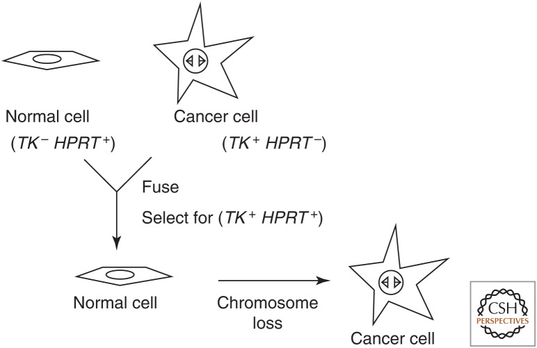
Evidence for tumor suppressor genes from cell fusion experiments. Variations of this idealized experiment used senescing normal cells or nonadherent cancer cells without their own genetic markers.
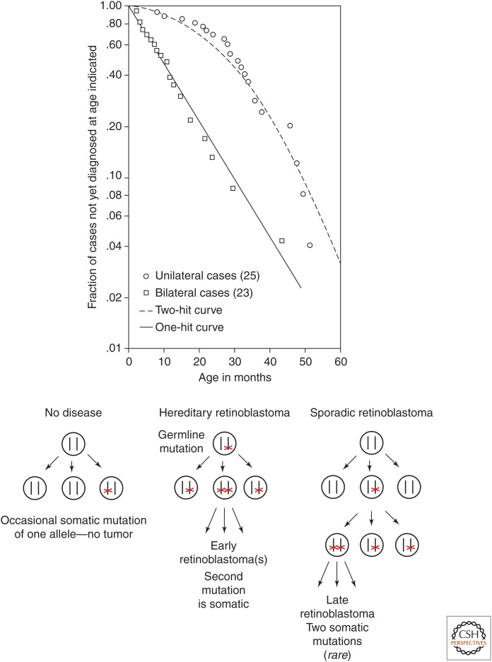
Knudson's two-hit hypothesis. (Top) Semilogarithmic plot of fraction of retinoblastoma cases not yet diagnosed versus age of patient. (Bottom) Model of events in hereditary versus sporadic retinoblastoma. (Top, Reprinted from Knudson AG. 1971. Proc Natl Acad Sci
68: 820–823, Fig. 1, p. 823.)
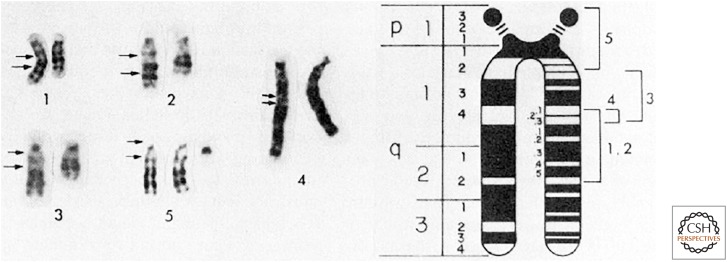
Deletions of chromosome 13 in patients with (1–4) or without (5) retinoblastoma. Arrows to the left of each normal chromosome indicate deleted or duplicated segments. The smallest region of overlapping deletions in retinoblastoma was in 13q14. (Reprinted from Sparkes RS, et al. 1980. Science
208: 1042–1044, Fig. 1, p. 1043, with permission from AAAS.)
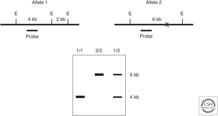
Genotyping using restriction fragment length polymorphism (RFLP). (Top) Cartoons of genomic DNA for two different alleles of the same locus. (E) EcoRI restriction enzyme recognition site, (X) absence of one EcoRI site in allele 2, (probe) radioactive DNA used for hybridization in the Southern blot below. (Bottom) Autoradiogram of a Southern blot of EcoRI-digested genomic DNA from individuals with the diploid genotypes indicated above each lane.
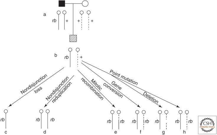
Mechanisms for generating somatic loss of heterozygosity (c–h) in retinoblasts of a heterozygous carrier (b) born to an affected father and unaffected mother (a). Solid lines represent paternal chromosomes; dashed lines represent maternal chromosomes. (Reprinted from Cavenee, et al. 1983. Nature
305: 779–784, Fig. 1, p. 780, by permission of Springer Nature.)
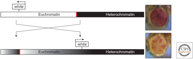
Position effect variegation of the white gene in Drosophila. The upper chromosome and eye are wild-type. The lower chromosome has an inversion that placed the white gene next to constitutive heterochromatin (black bar), resulting in a mottled eye. The red bar indicates a boundary that normally prevents the spread of silencing. (Reprinted from Elgin SC, Reuter G. 2013. Cold Spring Harb Perspect Biol
5: a017780, doi:10.1101/cshperspect.a017780, Fig. 1A, p. 4, © Cold Spring Harbor Laboratory Press.)
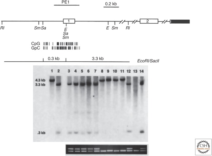
DNA methylation of the CDKN2A gene encoding p16INK4A in human lung cancer cell lines. (Top) Map of the locus showing restriction enzyme sites and the CpG island surrounding the first exon. (Middle) Southern blot with PE1 probe following digestion of genomic DNA with EcoRI (RI) and/or SacII (Sa). Lane 1 contains control DNA digested with EcoRI; lane 2 contains control DNA digested with EcoRI and SacII; lanes 3–14 contain DNAs from different lung cancer cell lines digested with EcoRI and SacII. Unmethylated DNA digested with EcoRI and SacII generates hybridizing fragments of 3.3 and 0.3 kb, whereas methylated DNA generates a hybridizing fragment of 4.0 kb because of the failure of SacII to cut at its recognition sites. (Bottom) RT-PCR detection of mRNA from the CDKN2A gene (top band) or a TP53 control gene (bottom band). Note the absence of CDKN2A mRNA in the cell lines in which the locus is methylated (lanes 8–11) or deleted (lane 13). (Adapted from Merlo et al. 1995. Nat Med
1: 686–682, Figs 1 and 2, pp. 687, 688, by permission of Springer Nature.)
Poster and T-shirt design for the 6th Annual Meeting on Oncogenes. (© Jamie Simon.)
References
-
- Baylin S, Bestor TH. 2002. Altered methylation patterns in cancer cell genomes: Cause or consequence? Cancer Cell 1: 299–305. - PubMed
-
- Cavenee WK, Dryja TP, Phillips RA, Benedict WF, Godbout R, Gallie BL, Murphree AL, Strong LC, White RL. 1983. Expression of recessive alleles by chromosomal mechanisms in retinoblastoma. Nature 305: 779–784. - PubMed
-
- Friend SH, Bernards R, Rogelj S, Weinberg RA, Rapaport JM, Albert DM, Dryja TP. 1986. A human DNA segment with properties of the gene that predisposes to retinoblastoma and osteosarcoma. Nature 323: 643–646. - PubMed
-
- Harris H. 1993. How tumour suppressor genes were discovered. FASEB J 7: 978–979. - PubMed
-
- Knudson AG. 2001. Two genetic hits (more or less) to cancer. Nat Rev Cancer 1: 157–162. - PubMed
Publication types
MeSH terms
LinkOut - more resources
Full Text Sources