. 2020 Feb 18;117(7):3779-3788.
doi: 10.1073/pnas.1915996117. Epub 2020 Feb 3.

Facilitative and synergistic interactions between fungal and plant viruses

Affiliations

Facilitative and synergistic interactions between fungal and plant viruses

Ruiling Bian et al. Proc Natl Acad Sci U S A. .

Abstract

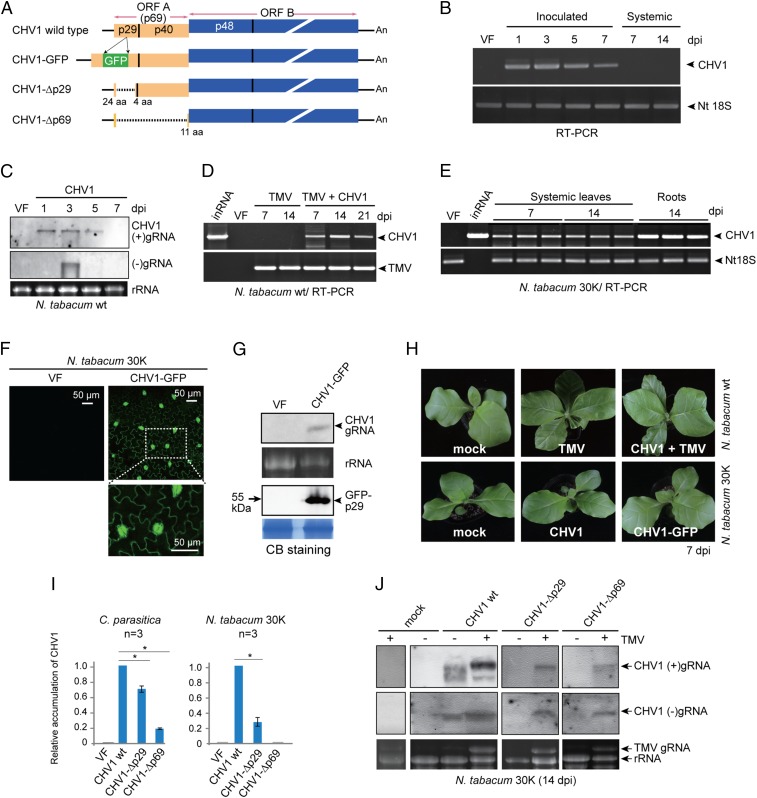
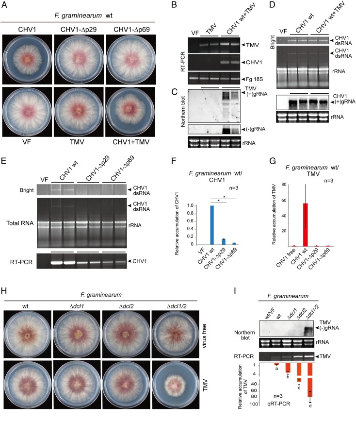
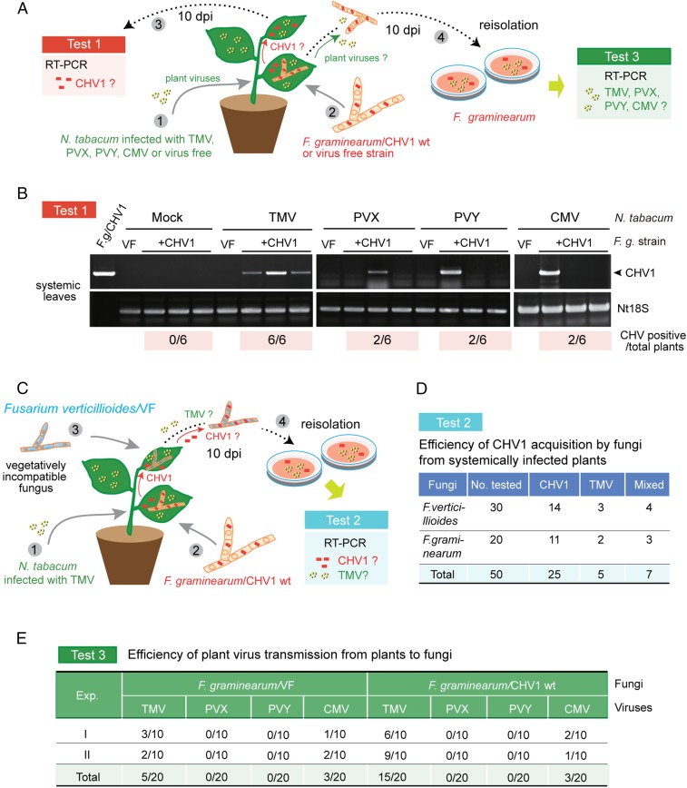
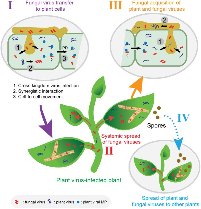
Plants and fungi are closely associated through parasitic or symbiotic relationships in which bidirectional exchanges of cellular contents occur. Recently, a plant virus was shown to be transmitted from a plant to a fungus, but it is unknown whether fungal viruses can also cross host barriers and spread to plants. In this study, we investigated the infectivity of Cryphonectria hypovirus 1 (CHV1, family Hypoviridae), a capsidless, positive-sense (+), single-stranded RNA (ssRNA) fungal virus in a model plant, Nicotiana tabacum CHV1 replicated in mechanically inoculated leaves but did not spread systemically, but coinoculation with an unrelated plant (+)ssRNA virus, tobacco mosaic virus (TMV, family Virgaviridae), or other plant RNA viruses, enabled CHV1 to systemically infect the plant. Likewise, CHV1 systemically infected transgenic plants expressing the TMV movement protein, and coinfection with TMV further enhanced CHV1 accumulation in these plants. Conversely, CHV1 infection increased TMV accumulation when TMV was introduced into a plant pathogenic fungus, Fusarium graminearum In the in planta F. graminearum inoculation experiment, we demonstrated that TMV infection of either the plant or the fungus enabled the horizontal transfer of CHV1 from the fungus to the plant, whereas CHV1 infection enhanced fungal acquisition of TMV. Our results demonstrate two-way facilitative interactions between the plant and fungal viruses that promote cross-kingdom virus infections and suggest the presence of plant-fungal-mediated routes for dissemination of fungal and plant viruses in nature.

Keywords: cross-kingdom; infection; mycovirus; plant virus.


Conflict of interest statement

The authors declare no competing interest.

Figures

Fig. 1.
Fig. 1.
Infection of CHV1 in N. tabacum plants. (A) Schematic presentation of the CHV1 WT, CHV1-GFP, CHV1-Δp29, and CHV1-Δp69 genomes (not to scale). For CHV1-GFP, the gfp gene was inserted into the p29 gene. Colored boxes, lines, and dashed lines indicate the ORF, untranslated regions (UTRs), and deleted regions, respectively. (B) RT-PCR detection of CHV1 gRNA accumulation in inoculated and upper uninoculated leaves of N. tabacum plants. (C) RNA blotting analysis of CHV1 (+) and (–)gRNA accumulation in inoculated leaves. VF, virus free. The RNA gel was stained with ethidium bromide, and rRNA is shown as a loading control. (D) RT-PCR detection of viral gRNA accumulation in the upper leaves of plants inoculated with TMV alone or with TMV plus CHV1. inRNA, in vitro CHV1 transcripts. (E) RT-PCR detection of CHV1 RNA accumulation in uninoculated upper leaves and roots of transgenic N. tabacum plants expressing the TMV 30K movement protein. (F) Fluorescence microscopy observations of GFP expression in epidermal cells of CHV1-GFP–infected upper uninoculated leaves. (G) RNA and Western blot analyses of CHV-GFP (+)gRNA and GFP-p29 fusion protein accumulation in CHV1-GFP–infected upper uninoculated leaves. Coomassie brilliant blue (CB)-stained total proteins run on separate gels with the same sample batch are shown as loading controls. (H) Phenotypic growth of N. tabacum inoculated with TMV alone or with TMV plus CHV1, and transgenic N. tabacum 30K plants infected with CHV1 or CHV1-GFP. (I) Quantitative RT-PCR analysis of gRNA accumulation of CHV1 WT and CHV1 mutants in C. parasitica and in upper uninoculated N. tabacum 30K leaves. “*” indicates significant differences (P < 0.05, Student’s t test). (J) RNA blotting analysis of CHV1 (+) and (–)gRNA accumulation in N. tabacum 30K plants infected with TMV.
Fig. 2.
Fig. 2.
CHV1 and TMV RNA accumulation in F. graminearum. (A) Phenotypic growth of fungal colonies that were virus-free (VF) or infected with WT CHV1, CHV1-Δp29, CHV1-Δp69, TMV, or CHV1 WT plus TMV. Fungi were grown on PDA medium (10-cm plates) for 3 d and then photographed. (B) RT-PCR detection of CHV1 and TMV RNA accumulation. (C) RNA blotting analysis of TMV (+) and (–)gRNA accumulation. (D) CHV1 dsRNA and (+)gRNA accumulation by gel electrophoresis and RNA blotting analyses. (E) dsRNA accumulation and RT-PCR detection of CHV1 WT and CHV1 mutants. (F) qRT-PCR analysis of CHV1 and CHV1 mutant RNA accumulation in F. graminearum. “*”indicates significant differences (P < 0.05, Student’s t test). (G) qRT-PCR analysis of TMV RNA accumulation in F. graminearum coinfected with CHV1 WT or CHV1 mutants. (H) Phenotypic growth of F. graminearum dcl mutant (Δdcl1, Δdcl2, and Δdcl1/2) fungal colonies in virus-free fungi or fungi infected with TMV. Fungi were grown on PDA medium (10-cm plates) for 3 d and then photographed. (I) RNA blotting and qRT-PCR analyses of TMV gRNA accumulation in F. graminearum dcl mutants. Different letters indicate significant differences (P < 0.05, one-way ANOVA).
Fig. 3.
Fig. 3.
Transmission of CHV1 from fungi to plants with the help of plant virus. (A) Experimental procedure for investigating horizontal transfer of CHV1 from F. graminearum to N. tabacum plants infected with different plant viruses (test 1) and acquisition of plant viruses by F. graminearum (test 3). (B) RT-PCR detection of CHV1 RNA accumulation in uninoculated upper leaves after systemic infection with TMV, PVX, PVY, CMV, or virus-free plants, as described in A (test 1). The numbers of CHV1-positive plants out of six tested plants are shown below the gel images. (C) Experimental procedure for investigating CHV1 acquisition by vegetatively incompatible F. verticillioides from TMV-infected plants systemically infected with CHV1 (test 2) after colonization by CHVI-carrying F. graminearum. (D) Efficiency of CHV1 and TMV acquisition by F. verticillioides or F. graminearum in the experiment described in C (test 2). (E) Efficiency of TMV, PVX, PVY, and CMV acquisition by F. graminearum infected with CHV1 or a virus-free fungal isolate in the experiment described in A (test 3). Viruses were detected by RT-PCR.
Fig. 4.
Fig. 4.
A proposed model for plant virus-facilitated spread of fungal viruses in nature. A fungal introduction of fungal viruses to plant cells during colonization of plant tissue (I). Fungal virus utilization of plant virus MPs for cell-to-cell and systemic spread in plants (II). Fungal virus acquisition by similar or different fungal species that concurrently colonize the plant. At the same time, the fungus may also acquire the plant virus (III). Fungal or plant virus vertical transmission to spores, and spread of fungal infections to other plants by virus infected spores (IV). These mechanisms permit fungal viruses to spread to vegetatively incompatible fungal strains or to different fungal species. In addition, fungi can also be plant virus vectors.

Similar articles

Cited by

References

    1. Ghabrial S. A., Suzuki N., Viruses of plant pathogenic fungi. Annu. Rev. Phytopathol. 47, 353–384 (2009). - PubMed
    1. Pearson M. N., Beever R. E., Boine B., Arthur K., Mycoviruses of filamentous fungi and their relevance to plant pathology. Mol. Plant Pathol. 10, 115–128 (2009). - PMC - PubMed
    1. Ghabrial S. A., Castón J. R., Jiang D., Nibert M. L., Suzuki N., 50-plus years of fungal viruses. Virology 479–480, 356–368 (2015). - PubMed
    1. King A. M. Q., et al. , Changes to taxonomy and the international code of virus classification and nomenclature ratified by the International Committee on Taxonomy of Viruses (2018). Arch. Virol. 163, 2601–2631 (2018). - PubMed
    1. Hillman B. I., Annisa A., Suzuki N., “Viruses of plant-interacting fungi” in Advances in Virus Research, Kielian M., Mettenleiter T. C., M. J. R., Eds. (Elsevier, 2018), vol. 100, pp. 99–116. - PubMed

Publication types

MeSH terms

Supplementary concepts

LinkOut - more resources