. 2020 Feb 18;117(7):3738-3747.
doi: 10.1073/pnas.1916588117. Epub 2020 Feb 3.

NAD deficiency due to environmental factors or gene-environment interactions causes congenital malformations and miscarriage in mice

Affiliations

NAD deficiency due to environmental factors or gene-environment interactions causes congenital malformations and miscarriage in mice

Hartmut Cuny et al. Proc Natl Acad Sci U S A. .

Abstract

Causes for miscarriages and congenital malformations can be genetic, environmental, or a combination of both. Genetic variants, hypoxia, malnutrition, or other factors individually may not affect embryo development, however, they may do so collectively. Biallelic loss-of-function variants in HAAO or KYNU, two genes of the nicotinamide adenine dinucleotide (NAD) synthesis pathway, are causative of congenital malformation and miscarriage in humans and mice. The variants affect normal embryonic development by disrupting the synthesis of NAD, a key factor in multiple biological processes, from its dietary precursor tryptophan, resulting in NAD deficiency. This study demonstrates that congenital malformations caused by NAD deficiency can occur independent of genetic disruption of NAD biosynthesis. C57BL/6J wild-type mice had offspring exhibiting similar malformations when their supply of the NAD precursors tryptophan and vitamin B3 in the diet was restricted during pregnancy. When the dietary undersupply was combined with a maternal heterozygous variant in Haao, which alone does not cause NAD deficiency or malformations, the incidence of embryo loss and malformations was significantly higher, suggesting a gene-environment interaction. Maternal and embryonic NAD levels were deficient. Mild hypoxia as an additional factor exacerbated the embryo outcome. Our data show that NAD deficiency as a cause of embryo loss and congenital malformation is not restricted to the rare cases of biallelic mutations in NAD synthesis pathway genes. Instead, monoallelic genetic variants and environmental factors can result in similar outcomes. The results expand our understanding of the causes of congenital malformations and the importance of sufficient NAD precursor consumption during pregnancy.

Keywords: NAD; congenital malformation; embryonic development; metabolism; miscarriage.


Conflict of interest statement

The authors declare no competing interest.

Figures

Fig. 1.
Fig. 1.
Phenotypic embryo outcomes at E18.5 in offspring from C57BL/6J wild-type mice show that, with a reduction of NAD precursors throughout pregnancy, the incidence of dead and malformed embryos increases. Mild hypoxia as an additional environmental factor leads to a further increase in the proportion of malformed embryos. (A) Percentages of normal embryos (green), embryos with isolated malformations (orange), embryos with more than one malformation (red), and dead embryos (black) within maternal diet treatment groups as indicated on the right. In this set of experiments, all mice were Haao+/+. Table 2 shows statistics and embryo numbers. M, maternal Haao genotype; P, paternal Haao genotype. (B) Incidence of specific organ defects. Black bars represent percentages of embryos among the respective treatment group that exhibited the indicated defect. Lines and asterisks indicate statistical comparison of malformation incidence between standard, NTF + TW600, and NTF + TW500 groups (light blue) by Fisher’s exact test with Freeman–Halton extension and NTF + TW600 and NTF + TW600 + HYP groups (orange) by two-sided Fisher’s exact test. The vertebrae category includes rib malformations. Abd. wall, abdominal wall; ns, not significant; X, malformation types observed in isolation in one or more embryos. *P < 0.05; ****P < 0.0001.
Fig. 2.
Fig. 2.
Phenotypic embryo outcomes at E18.5 in offspring from mice with an Haao+/− mutation show a high incidence of embryo mortality with NTF + TW600 diet and indicate a gene–environment effect. With additional supply of NA in the water (NTF + TW600 + NW15), embryo mortality is reduced. (A) Summarized embryo outcomes. The dietary treatments throughout pregnancy are indicated on the right. Table 2 shows statistics and embryo numbers. M, maternal Haao genotype; P, paternal Haao genotype. (B) Incidence of specific organ defects occurring in the respective treatment condition (Left) and of the different embryo Haao genotypes within the NTF + TW600 group (Right). Asterisks indicate significant differences between the three embryo Haao genotypes by Fisher’s exact test with Freeman–Halton extension. The vertebrae category includes rib malformations. Abd. wall, abdominal wall; ns, not significant; X, malformation types observed in isolation in one or more embryos. *P < 0.05; **P < 0.01; ***P < 0.001; ****P < 0.0001.
Fig. 3.
Fig. 3.
Maternal liver and embryo total NAD levels (NAD+ and NADH) and phenotypic embryo outcomes at E11.5. Dots represent NAD levels, and bars indicate the mean ± SD. (A) Maternal liver NAD levels. Treatment groups are indicated on the top. The parental and embryo Haao genotypes are indicated at the bottom. Liver tissue was collected at E11.5 along with the embryos. The first column represents liver NAD levels of nonpregnant females maintained on standard diet for at least 3 wk prior to dissection, which were of similar age to the pregnant females. (B) Whole-embryo NAD levels. Analyzed embryos were offspring of the mothers with liver NAD levels that were measured. The total numbers and percentages of live and dead embryos observed with each treatment condition are indicated at the bottom. Note that not every collected embryo underwent NAD measurement. Asterisks indicate NAD levels that are significantly different to those of the pregnant C57BL/6J wild-type standard diet group (second column) by one-way ANOVA with Dunnett’s multiple comparisons test with *P < 0.05; **P < 0.01; ***P < 0.001; ****P < 0.0001. A summary of NAD level values is in Table 3 and SI Appendix, Table S3. n.a., not applicable (no embryos in nonpregnant females); ns, not significant.
Fig. 4.
Fig. 4.
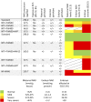
Summary of NAD levels and phenotypic outcomes of all treatment conditions tested in this study. Four thresholds for maternal liver and embryo NAD levels were set to specify normal, mild, severe, and very severe NAD deficiency. Similarly, four levels for the incidence of affected embryos at E18.5 were set. For the classification of embryo outcomes at E18.5 involving Haao genotypes, the assumption was made that the genotypes of dead (resorbed) embryos, which could not be genotyped, were distributed in a normal Mendelian ratio (25:50:25) based on the finding that the genotypes of surviving embryos did not significantly deviate from a normal Mendelian ratio.

Comment in

Similar articles

Cited by

References

    1. Hobbs C. A., et al. , Genetic epidemiology and nonsyndromic structural birth defects: From candidate genes to epigenetics. JAMA Pediatr. 168, 371–377 (2014). - PMC - PubMed
    1. Rai R., Regan L., Recurrent miscarriage. Lancet 368, 601–611 (2006). - PubMed
    1. Kaiser J., Branch D. W., Recurrent pregnancy loss: Generally accepted causes and their management. Clin. Obstet. Gynecol. 59, 464–473 (2016). - PubMed
    1. Baldacci S., et al. , Environmental and individual exposure and the risk of congenital anomalies: A review of recent epidemiological evidence. Epidemiol. Prev. 42 (suppl. 1), 1–34 (2018). - PubMed
    1. Zhu H., Kartiko S., Finnell R. H., Importance of gene-environment interactions in the etiology of selected birth defects. Clin. Genet. 75, 409–423 (2009). - PubMed

Publication types

LinkOut - more resources