. 2020 Feb 3;12(2):025002.
doi: 10.1088/1758-5090/ab6413.

Handheld instrument for wound-conformal delivery of skin precursor sheets improves healing in full-thickness burns

Affiliations

Handheld instrument for wound-conformal delivery of skin precursor sheets improves healing in full-thickness burns

Richard Y Cheng et al. Biofabrication. .

Abstract

The current standard of care for patients with severe large-area burns consists of autologous skin grafting or acellular dermal substitutes. While emerging options to accelerate wound healing involve treatment with allogeneic or autologous cells, delivering cells to clinically relevant wound topologies, orientations, and sizes remains a challenge. Here, we report the one-step in situ formation of cell-containing biomaterial sheets using a handheld instrument that accommodates the topography of the wound. In an approach that maintained cell viability and proliferation, we demonstrated conformal delivery to surfaces that were inclined up to 45° with respect to the horizontal. In porcine pre-clinical models of full-thickness burn, we delivered mesenchymal stem/stromal cell-containing fibrin sheets directly to the wound bed, improving re-epithelialization, dermal cell repopulation, and neovascularization, indicating that this device could be introduced in a clinical setting improving dermal and epidermal regeneration.


Figures

Figure 1.
Figure 1.. In-situ formation of precursor skin tissue using an intraoperative approach.
a, Schematic illustration of handheld approach for delivering cell-laden biomaterial sheets conformal to full-thickness burn wound of arbitrary size and topology. b, Rendered image of handheld instrument for controllable delivery of bioink consisting of mesenchymal stem/stromal cells (MSCs) in fibrin-based bioink (green color) supplied at flow rate QB with cross-linker (clear) supplied at flow rate QC through microfluidic printhead, while driven by soft wheel along wound surface at velocity V.
Figure 2.
Figure 2.. Conformal deposition of biomaterial layers onto physiologically relevant topologies.
a, 3D rendering, exploded view of handheld instrument and disposable bioink syringes, microfluidic printhead, and silicone wheel. b, Left: Side view of printhead illustrating conformal sheet deposition by printhead onto wound substrate being unaffected by wheel deformation. Right: contact pressures measured for wheels of different hardness on stiff and soft substrates. Dotted line corresponds to stiffness of wound tissue. Data expressed as mean ± s.d., n = 5 independent experiments. c, Left: rendered image of printhead rotating about y-axis with pitch angle α, compensating for inclinations of up to 45°. Right: printhead rotation about x-axis with roll angle φ, accounting for ±25° variation in instrument position with respect to the normal direction of the deposition surface. d, Left: side view photograph of bioink extrusion. QC and QB indicate perfusion of cross-linker and bioink through printhead (Supplementary Table 1; right column). Wheel rotates clockwise to advance instrument at nominal speed V0 in deposition direction. Middle: schematic cross-sectional view showing bioink and cross-linker exiting printhead to form biomaterial sheets on deposition surface with height hT. Right: photograph of fibrin sheet formation. Scale bar: 2mm. e, Micrographs of deposited sheets for different values of V0 and total flow rate Q and expected sheet width w for conditions of uniform coverage (top), sheet contraction (middle), and non-uniform coverage (bottom). Scale bar: 10mm. f, Top: percentage area covered for V0 = 3mm/s. Bottom: measured actual wheel speed, V, versus V0. Data expressed as mean ± s.d., n = 3 independent experiments; *p<0.05, t-test. g, Graphical representation of operating conditions to generate biomaterial sheets of varying thicknesses depending on total flow rate (Q) and wheel speed (V0) for expected sheet width (w). h, Projected time, biomaterial volume, crosslinker volume, and cell number required for area coverage for hT = 0.2mm, c = 1×10⁶ cells/ml, and w = 25mm or 50mm. Blue dots: experimental conditions used in vivo, n = 9 independent wounds.
Fig. 3.
Fig. 3.. Fibrin-HA biomaterials maintain uniform coverage over tilted surfaces following microfluidic extrusion to provide a homogenous environment for 3D cell growth.
a, Schematic of biomaterial sheet behavior after deposition from microfluidic printhead (Supplementary Table 1; right side) onto surface with inclination angle Θ. Selected values represent inclination angles of burn wounds in vivo. Biomaterial sheet of initial width w undergoes drainage-induced reduction in thickness, Δh, and lateral translation, Δy. b, Viscosity measured at different shear rates and hyaluronic acid content in fibrinogen-based bioink. c, Time tg associated with reaching 95% change in turbidity as obtained through confocal microscopy. Data expressed as mean ± s.d., n = 3 independent experiments. d, Drainage velocity as determined by tracking 1% HA-containing biomaterial sheets containing 1μm microspheres on surface with tilt angle Θ. Data expressed as mean ± s.d., n = 5 individually tracked particles. Black lines indicate best fit, R2 > 0.91. e, Left: time ts until drainage ended due to gelation-induced viscosity increase. Center: Relative lateral drainage distance Δy, at time ts. Right: percentage change in biomaterial thickness Δh due to drainage at time ts determined from confocal microscopy. f, Representative time snaps of 1×10 MSC/ml fibrin-HA biomaterials in 3D culture for 0 to 7 days after sheet deposition, stained for cell nucleus (Hoechst, blue) and α-Actin (Phalloidin, green). Scale bar: 100μm. g, Quantification of cell viability, cell number, distance to nearest neighbour, and cell aspect ratio of MSCs in 3D culture after microfluidic extrusion from days 0 to 7. Data expressed as mean ± s.d., n = 3 independent experiments.
Fig. 4.
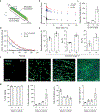
Fig. 4.. MSC-containing fibrin-HA biomaterials deposited homogenously on a porcine full thickness burn surface using the handheld device contribute to improved wound healing.
a, Left: photograph showing the side view of the handheld device in the process of depositing MSC-containing fibrin-HA biomaterials onto a porcine full-thickness burn model with only the microfluidic printhead and silicone wheel in contact with the wound substrate. Right: isometric view of the handheld instrument during the deposition process, with the body of the instrument positioned at a different angle relative to the microfluidic printhead, which is held parallel and in direct contact with the wound substrate underneath. Scale bar: 2.5cm b, Top down photographs showing the 5cm × 5cm burned wound prior to (left) and immediately after (right) in-situ deposition. Scale bar: 2cm c, Top down photographs indicating macroscopic wound healing after 28 days of recovery comparing acellular biomaterial treatment only (left), and MSC-containing biomaterials (right). Scale bar: 2cm. d, Masson`s Trichrome stained tissue sections of healthy skin and wounds treated with burn alone, acellular biomaterials only, and MSC-containing biomaterials after 28 days post-biomaterial deposition. Scale bar: 200μm. e, Quantification of epithelialization speed, scar quality, and contracture per wound from macroscopic assessment. f, Quantification of epidermal thickness, collagen density, and CD31+ vessels from histology. g, Quantification of CD163+ expression, CD11b+ expression, and a-SMA+ density from histology. Data expressed as median IQR, n = 4 separate pig models with >3 replicates per condition per animal, *p<0.05, Wilcoxon–Mann–Whitney test.

References

    1. Moiemen N, Lee K and Joory K 2014. History of burns: The past, present and the future Burn. Trauma 2 169 - PMC - PubMed
    1. Chua A W C, Khoo Y C, Tan B K, Tan K C, Foo C L and Chong S J 2016. Skin tissue engineering advances in severe burns: review and therapeutic applications Burn. Trauma 4 - PMC - PubMed
    1. Rosanova M T, Stamboulian D and Lede R 2014. Risk factors for mortality in burn children Brazilian J. Infect. Dis. 18 144–9 - PMC - PubMed
    1. Greenhalgh D G, Saffle J R, Holmes J H, Gamelli R L, Palmieri T L, Horton J W, Tompkins R G, Traber D L, Mozingo D W, Deitch E A, Goodwin C W, Herndon D N, Gallagher J J, Sanford A P, Jeng J C, Ahrenholz D H, Neely A N, O’Mara M S, Wolf S E, Purdue G F, Garner W L, Yowler C J and Latenser B A 2007. American burn association consensus conference to define sepsis and infection in burns Journal of Burn Care and Research vol 28 pp 776–90 - PubMed
    1. Galeiras R, Lorente J A, Pértega S, Vallejo A, Tomicic V, de la Cal M A, Pita S, Cerdá E and Esteban A 2009. A model for predicting mortality among critically ill burn victims Burns 35 201–9 - PubMed

Publication types

MeSH terms