

Ultrastructural and immunohistochemical characteristics of telocytes in human scalp tissue
- PMID: 32015359
- PMCID: PMC6997163
- DOI: 10.1038/s41598-020-58628-w
Ultrastructural and immunohistochemical characteristics of telocytes in human scalp tissue
Abstract
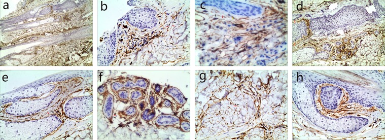
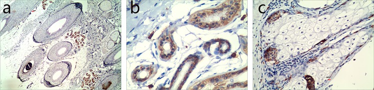
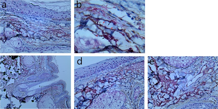
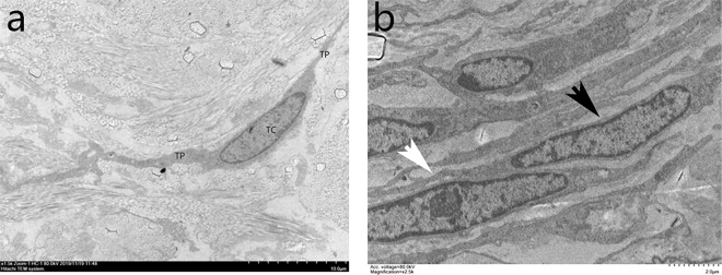
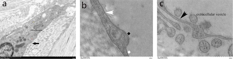
This study was designed to characterize the location, morphology and ultrastructure of telocytes (TCs) in human scalp tissue. After obtaining approval for this study and informed consent from the patient, a scalp specimen was obtained. The distribution and morphology of TCs in human scalp tissue was assessed by immunohistochemical staining of CD34 and CD117/c-KIT, and the ultrastructure of TCs was investigated using transmission electron microscopy (TEM). Immunohistochemical staining of CD34 revealed that TCs were located in the connective tissue of human scalp, and were concentrated around hair follicles (HFs), blood vessels, sweat glands, sebaceous glands and adipose lobules. Immunohistochemical staining of CD117 revealed that TCs were mainly located in the dermis of human scalp, surrounding the HFs and sweat glands. Under TEM, TCs were seen and confirmed by their special morphological features. These cells were spindle-shaped, had small cell bodies and long thin processes, and surrounded stem cell clusters in the bulge region of HFs. These results demonstrate that TCs in human scalp were positive for CD34 and CD117, and their strategic positioning surrounding stem cells suggests their possible involvement in local regeneration, remodeling and homeostasis of the skin.
Conflict of interest statement
The authors declare no competing interests.
Figures
Similar articles
-
Telocytes in human skin--are they involved in skin regeneration?J Cell Mol Med. 2012 Jul;16(7):1405-20. doi: 10.1111/j.1582-4934.2012.01580.x. J Cell Mol Med. 2012. PMID: 22500885 Free PMC article.
-
Localization of telocytes in rabbits testis: Histological and immunohistochemical approach.Microsc Res Tech. 2018 Nov;81(11):1268-1274. doi: 10.1002/jemt.23133. Epub 2018 Oct 23. Microsc Res Tech. 2018. PMID: 30351479
-
Immunohistochemical and ultrastructural evidence for telocytes in the different physiological stages of the female rat mammary gland.Life Sci. 2019 Aug 15;231:116521. doi: 10.1016/j.lfs.2019.05.077. Epub 2019 May 29. Life Sci. 2019. PMID: 31152814
-
The Cutaneous Telocytes.Adv Exp Med Biol. 2016;913:303-323. doi: 10.1007/978-981-10-1061-3_20. Adv Exp Med Biol. 2016. PMID: 27796896 Review.
-
Hepatic Telocytes.Adv Exp Med Biol. 2016;913:425-432. doi: 10.1007/978-981-10-1061-3_27. Adv Exp Med Biol. 2016. PMID: 27796903 Review.
Cited by
-
Analysis of TP53 gene and particular infrastructural alterations in invasive ductal mammary carcinoma.Rom J Morphol Embryol. 2020 Apr-Jun;61(2):441-447. doi: 10.47162/RJME.61.2.13. Rom J Morphol Embryol. 2020. PMID: 33544795 Free PMC article.
-
Cd34+ Stromal Cells/Telocytes in Normal and Pathological Skin.Int J Mol Sci. 2021 Jul 8;22(14):7342. doi: 10.3390/ijms22147342. Int J Mol Sci. 2021. PMID: 34298962 Free PMC article. Review.
-
Immunohistochemical and Ultrastructural Characterization of Telocytes in Normal and Diabetic Human Kidneys.Biomolecules. 2024 Aug 8;14(8):968. doi: 10.3390/biom14080968. Biomolecules. 2024. PMID: 39199356 Free PMC article.
-
Telocytes and their structural relationships with surrounding cell types in the skin of silky fowl by immunohistochemistrical, transmission electron microscopical and morphometric analysis.Poult Sci. 2021 Sep;100(9):101367. doi: 10.1016/j.psj.2021.101367. Epub 2021 Jun 29. Poult Sci. 2021. PMID: 34325111 Free PMC article.
-
Delimiting CD34+ Stromal Cells/Telocytes Are Resident Mesenchymal Cells That Participate in Neovessel Formation in Skin Kaposi Sarcoma.Int J Mol Sci. 2023 Feb 14;24(4):3793. doi: 10.3390/ijms24043793. Int J Mol Sci. 2023. PMID: 36835203 Free PMC article.
References
Publication types
MeSH terms
Substances
LinkOut - more resources
Full Text Sources
