. 2020 Feb 3;10(1):1667.
doi: 10.1038/s41598-020-58548-9.

Photodynamic therapy in oral lichen planus: A prospective case-controlled pilot study

Affiliations

Photodynamic therapy in oral lichen planus: A prospective case-controlled pilot study

Raluca Cosgarea et al. Sci Rep. .

Abstract

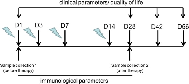
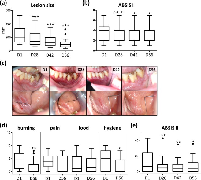
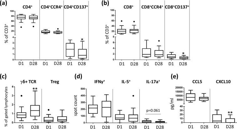
Oral lichen planus (OLP) is a common, chronic relapsing inflammatory disorder of the mucous membranes, which causes major discomfort. Current treatment includes topical/systemic glucocorticoids, immune modulators and systemic immunosuppressants, which may lead to considerable side-effects. The aim of this study was to determine the clinical and immunological efficacy of photodynamic therapy (PDT) in OLP as an alternative, easy-to-use, safe and non-invasive treatment. Twenty patients with OLP were treated with PDT in a prospective case-controlled pilot-study. PDT was performed on the most extensive oral lesion in 4 sessions (day 1, 3, 7, 14). Peripheral blood and lesional T cells were analysed before (day 1) and after PDT treatment (day 28). PDT led to a statistically significant reduction of clinical parameters (lesion size, ABSIS, Thongprasom-score) and improvement of all evaluated quality-of-life (QOL) items. The clinical improvement was accompanied by a significant decrease of the relative number of CD4+ and CD8+ T cells in mucosal OLP-lesions. Furthermore, CXCL10 plasma levels were decreased and the number of activated peripheral CD4 + CD137+ and CD8 + CD137+ T cells and IL-17-secreting T cells was diminished. PDT treatment in OLP leads to lesion reduction and improvement of QOL, and induces local and systemic anti-inflammatory effects. The study identifies PDT as a novel therapeutic option in OLP.


Conflict of interest statement

The authors declare no competing interests.

Figures

Figure 1
Figure 1
Time line of PDT treatment. Clinical and QOL parameters were assessed on day 1 (D1; before therapy), day 28 (D28, 14 days after therapy), day 42 (D42, 28 days after therapy) and day 56 (D56; 42 days after therapy). Samples were collected at D1 and D28. PDT, photodynamic treatment; QOL, quality of life.
Figure 2
Figure 2
PDT leads to clinical amelioration of OLP. (a) PDT induces a reduction of lesion size over time (D1-D56). (b) PDT leads to reduction of ABSIS score I over time (D1-D56). (c) Photographically documentation of lesion size reduction over time (D1-D56). (d) PDT leads to a reduction of ABSIS II score over time (D1-D56). (e) QoL parameters (burning, pain, food, oral hygiene) are improved after PDT treatment (D1-D56). *p < 0.05, **p < 0.01, ***p < 0.001. PDT, photodynamic treatment; QOL, quality of life.
Figure 3
Figure 3
PDT induces systemic anti-inflammatory effects in OLP. (a) Relative number of CD137 expressing peripheral blood CD4+ T- cells is decreased 14 days after PDT treatment. Results based on analysis by flow cytometry. (b) Relative number of CD137 expressing peripheral blood CD8+ T- cells is decreased 14 days after PDT treatment. Results based on analysis by flow cytometry. (c) PDT leads to an increase of the relative number of peripheral blood γδ T cells. Results based on analysis by flow cytometry. (d) Number of peripheral blood IL-17a producing T cells is decreased 14 days after PDT treatment. Results based on ELISpot. analysis. (e) CXCL10 plasma levels are decreased 14 days after PDT treatment. Results based on ELISA analysis.*p < 0.05, **p < 0.01. PDT, photodynamic treatment.
Figure 4
Figure 4
CXCL8 saliva levels are slightly decreased after PDT treatment. Shown are saliva concentrations (pg/ml) of CXCL8 before (day 1, D1) and after (day 28, D28) PDT treatment of oral OLP lesions.
Figure 5
Figure 5
PDT induces local anti-inflammatory effects in OLP. (a) Overview of immunohistochemical staining of mucosal tissue sections with a lesional T- cell infiltrate before and after PDT treatment (D1, D28). (b–d) Relative number of lesional CD3+, CD4+ and CD8+ cells are decreased 14 days after PDT treatment. (e) Relative number of lesional IL-17+ cells is increased 14 days after PDT treatment. **p < 0.01. PDT, photodynamic treatment.
Figure 6
Figure 6
Analysis of T lymphocyte subsets in OLP lesions. The T- cell infiltrate of mucosal sections was quantified by microscopical images (Axiostar, Zeiss, Jena, Germany) in combination with Cell^D (Soft Imaging System, Berlin, Germany) and ImageJ software (freeware). T cells were counted at a 100x magnification. After generating a grid (ImageJ software; area per point: 50.000 pixels^2), all stained T- cells were counted in three squares adjacent to basal membrane zone (ImageJ, cell counter) and their proportion of all infiltrating cells was determined subsequently.

Similar articles

Cited by

References

    1. Lavanya N, Jayanthi P, Rao UK, Ranganathan K. Oral lichen planus: An update on pathogenesis and treatment. Journal of oral and maxillofacial pathology: JOMFP. 2011;15:127–132. doi: 10.4103/0973-029X.84474. - DOI - PMC - PubMed
    1. Lodi G, Carrozzo M, Furness S, Thongprasom K. Interventions for treating oral lichen planus: a systematic review. The British journal of dermatology. 2012;166:938–947. doi: 10.1111/j.1365-2133.2012.10821.x. - DOI - PubMed
    1. Lodi G, et al. Current controversies in oral lichen planus: report of an international consensus meeting. Part 1. Viral infections and etiopathogenesis. Oral surgery, oral medicine, oral pathology, oral radiology, and endodontics. 2005;100:40–51. doi: 10.1016/j.tripleo.2004.06.077. - DOI - PubMed
    1. Thornhill MH. Immune mechanisms in oral lichen planus. Acta odontologica Scandinavica. 2001;59:174–177. doi: 10.1080/000163501750266774. - DOI - PubMed
    1. Aghahosseini F, Arbabi-Kalati F, Fashtami LA, Fateh M, Djavid GE. Treatment of oral lichen planus with photodynamic therapy mediated methylene blue: a case report. Medicina oral, patologia oral y cirugia bucal. 2006;11:E126–9. - PubMed

Publication types

Substances