. 2020 Feb 3;10(1):1673.
doi: 10.1038/s41598-020-58612-4.

Quantitative spatio-temporal characterization of epileptic spikes using high density EEG: Differences between NREM sleep and REM sleep

Affiliations

Quantitative spatio-temporal characterization of epileptic spikes using high density EEG: Differences between NREM sleep and REM sleep

Xuan Kang et al. Sci Rep. .

Abstract

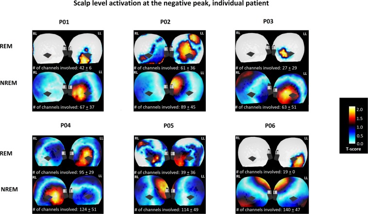
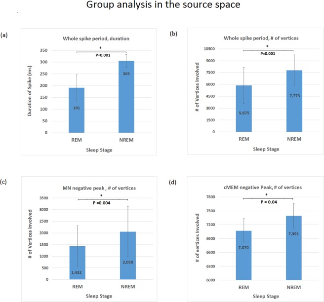
In this study, we applied high-density EEG recordings (HD-EEG) to quantitatively characterize the fine-grained spatiotemporal distribution of inter-ictal epileptiform discharges (IEDs) across different sleep stages. We quantified differences in spatial extent and duration of IEDs at the scalp and cortical levels using HD-EEG source-localization, during non-rapid eye movement (NREM) sleep and rapid eye movement (REM) sleep, in six medication-refractory focal epilepsy patients during epilepsy monitoring unit admission. Statistical analyses were performed at single subject level and group level across different sleep stages for duration and distribution of IEDs. Tests were corrected for multiple comparisons across all channels and time points. Compared to NREM sleep, IEDs during REM sleep were of significantly shorter duration and spatially more restricted. Compared to NREM sleep, IEDs location in REM sleep also showed a higher concordance with electrographic ictal onset zone from scalp EEG recording. This study supports the localizing value of REM IEDs over NREM IEDs and suggests that HD-EEG may be of clinical utility in epilepsy surgery work-up.


Conflict of interest statement

The authors declare no competing interests.

Figures

Figure 1
Figure 1
Group level analysis at the scalp level: asterisk is applied for result that reached statistical significance. (a) IED duration based on analysis of the whole spiking period, with shorter duration (p = 5 × 10−11) during REM sleep. (b) Number of channels involved during IED based on analysis of the whole spiking period, with less channels involved (p = 2 × 10−5) during REM sleep. (c) Number of channels involved at the negative peak, with less channels involved (p = 0.01) during REM sleep.
Figure 2
Figure 2
Graphic interpretation of IED scalp level analysis at negative peak. Each subject number is labeled on top of the corresponding figure. For each subject, the left and right lateral view of scalp level activation was demonstrated. The top row represents IED involvement during REM sleep, and the bottom row represents IED involvement during NREM sleep. RL = right lateral; LL = left lateral.
Figure 3
Figure 3
Source localization analysis of IED at different sleep stage: asterisk is applied for result that reached statistical significance. (a) IED duration based on source level analysis of the whole spiking period using MN estimate, with shorter duration (p = 0.001) during REM sleep. (b) Number of vertices on the cortex involved during IED based on source level analysis of the whole spiking period using MN Estimate, with less channels involved (P = 0.001) during REM sleep. (c) Number of vertices on the cortex involved at negative peak based on source level analysis using MN Estimate, with less channels involved (p = 0.005) during REM sleep. (d) Number of vertices on the cortex involved at negative peak based on source level analysis using cMEM Estimate, with less channels involved (p = 0.04) during REM sleep.
Figure 4
Figure 4
Graphic interpretation of IED source level analysis using cMEM at negative peak. Each subject number is labeled on top of the corresponding figure. For each subject, the left and right lateral view of the cortical reconstruction was demonstrated. The top row represents IED involvement during REM sleep, and the bottom row represents IED involvement during NREM sleep. Asterisk indicates IED with consistent localization as ictal recording obtained using 10–20 system during the same EMU stay. RL = right lateral; LL = left lateral.

Similar articles

Cited by

References

    1. Nevalainen O, et al. Epilepsy-related clinical characteristics and mortality: a systematic review and meta-analysis. Neurology. 2014;83:1968–77. doi: 10.1212/WNL.0000000000001005. - DOI - PubMed
    1. Kwan P, Sander JW. The natural history of epilepsy: an epidemiological view. J. Neurol. Neurosurg Psychiatry. 2004;75:1376–81. doi: 10.1136/jnnp.2004.045690. - DOI - PMC - PubMed
    1. Boly M, et al. Altered sleep homeostasis correlates with cognitive impairment in patients with focal epilepsy. Brain. 2002;140:1026–1040. doi: 10.1093/brain/awx017. - DOI - PMC - PubMed
    1. Van Schalkwijk FJ, et al. The impact of sleep characteristics and epilepsy variables on memory performance in patients with focal seizures. Epilepsy Behav. 2018;87:152–158. doi: 10.1016/j.yebeh.2018.06.034. - DOI - PubMed
    1. De Tisi J, et al. The long-term outcome of adult epilepsy surgery, patterns of seizure remission, and relapse: a cohort study. Lancet. 2011;378:1388–1395. doi: 10.1016/S0140-6736(11)60890-8. - DOI - PubMed

Publication types