. 2020 Feb 3;10(1):1707.
doi: 10.1038/s41598-020-58500-x.

Improved Calcium Homeostasis and Force by Selenium Treatment and Training in Aged Mouse Skeletal Muscle

Affiliations

Improved Calcium Homeostasis and Force by Selenium Treatment and Training in Aged Mouse Skeletal Muscle

János Fodor et al. Sci Rep. .

Abstract

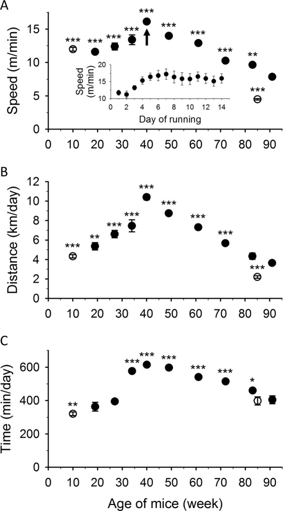
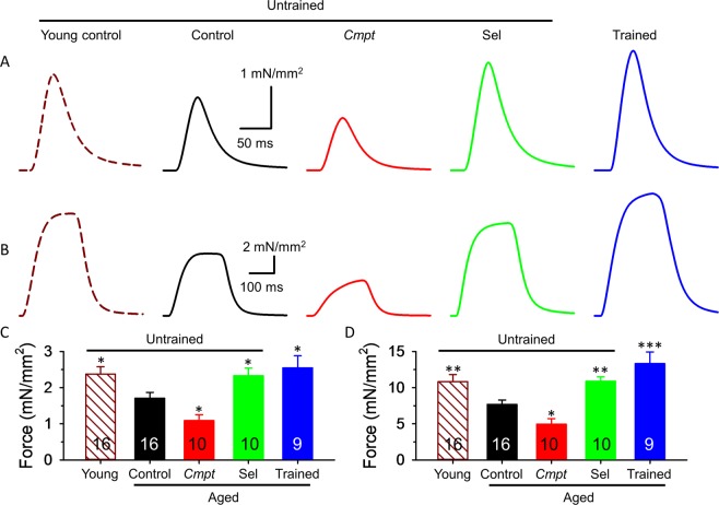
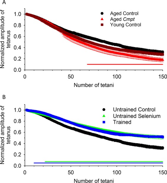
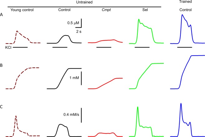
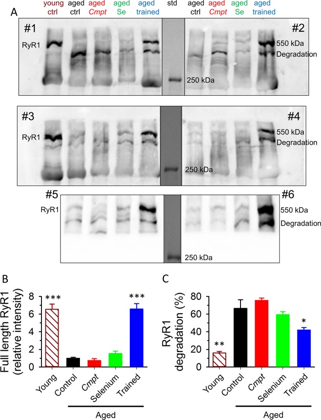
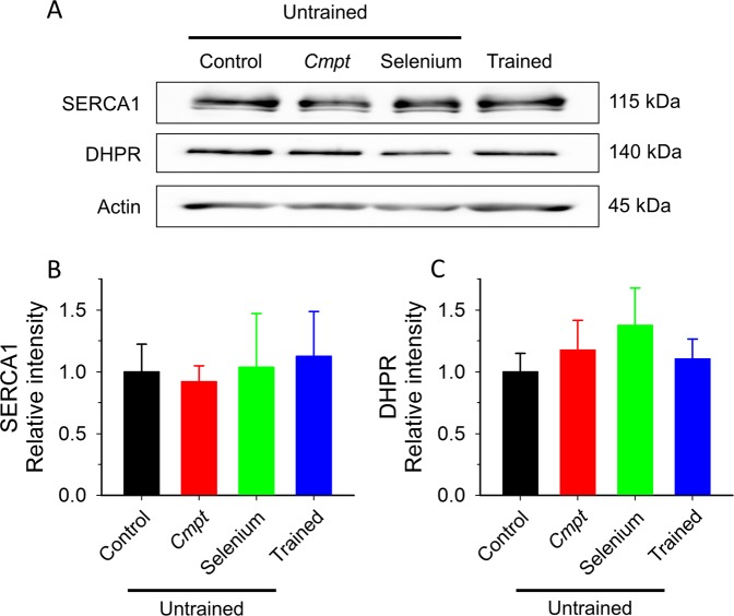
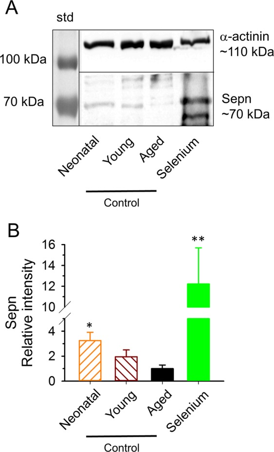
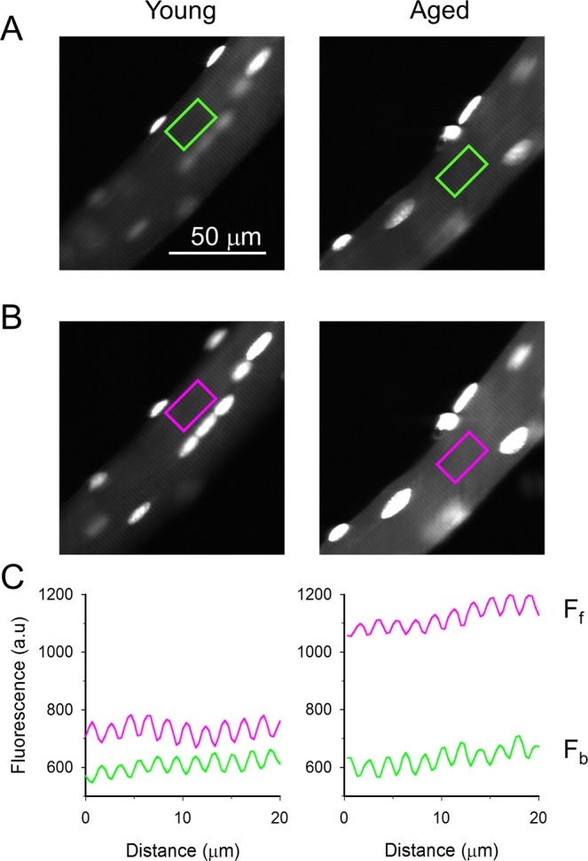
During aging reduction in muscle mass (sarcopenia) and decrease in physical activity lead to partial loss of muscle force and increased fatigability. Deficiency in the essential trace element selenium might augment these symptoms as it can cause muscle pain, fatigue, and proximal weakness. Average voluntary daily running, maximal twitch and tetanic force, and calcium release from the sarcoplasmic reticulum (SR) decreased while reactive oxygen species (ROS) production associated with tetanic contractions increased in aged - 22-month-old - as compared to young - 4-month-old - mice. These changes were accompanied by a decline in the ryanodine receptor type 1 (RyR1) and Selenoprotein N content and the increased amount of a degraded RyR1. Both lifelong training and selenium supplementation, but not the presence of an increased muscle mass at young age, were able to compensate for the reduction in muscle force and SR calcium release with age. Selenium supplementation was also able to significantly enhance the Selenoprotein N levels in aged mice. Our results describe, for the first time, the beneficial effects of selenium supplementation on calcium release from the SR and muscle force in old age while point out that increased muscle mass does not improve physical performance with aging.


Conflict of interest statement

The authors declare no competing interests.

Figures

Figure 1
Figure 1
Average speed, daily voluntary running distance and time. Average speed (A) daily distance (B) and time (C) of eight control mice during voluntary running for two weeks in every two months. Empty symbols represent data from measurements on young and aged mice which did not run previously. Inset in panel A shows a typical accommodation of a mouse to the new environment in the cage with the running wheel at the age marked with the arrow. *, **, and *** denote significant difference from the values at 90 weeks of trained aged animals at p < 0.05, p < 0.01, and p < 0.001, respectively.
Figure 2
Figure 2
Isometric force in EDL muscle. Twitch (A,C) and tetanus (B,D) in EDL muscles of 4-month-old untrained control (brown, dashed line), 20-month-old untrained control (black), Cmpt (red), selenium fed (green), and trained control (blue) mouse at room temperature (24 °C). The force was normalized to cross section of the muscle. Numbers in bars give the number of muscles investigated. *, **, and *** denote significant difference from control at p < 0.05, p < 0.01, and p < 0.001, respectively. The number of animals investigated is given in Table 2.
Figure 3
Figure 3
Fatigue measurement. Fatigue of EDL was evoked with 150 tetani at frequency of 0.5 Hz and the amplitude of consecutive tetani was normalized to the first tetanus developed. (A) Fatigue was similar in young and aged control mice but significantly higher in aged Cmpt than in the other mice. (B) Selenium and training increased fatigue resistance in aged mice. Solid horizontal lines below the data points (red: untrained Cmpt; green: untrained selenium fed; blue: trained control) represent the interval where data are significantly different from untrained aged control at p < 0.05. The number of muscles investigated is given in Table 2.
Figure 4
Figure 4
KCl depolarization-evoked Ca2+ transients and SR Ca2+ release in FDB fibers. Depolarization- (KCl) evoked changes in intracellular calcium concentration (A), amount of Ca2+ released (B), and Ca2+ flux (C) in FDB muscle fibers of 4-month-old untrained control (brown, dashed line), 20-month-old untrained control (black), Cmpt (red), selenium fed (green), and trained control (blue) mouse. Black horizontal lines below the calcium transients represent the application of KCl.
Figure 5
Figure 5
Expression of RyR1 in aged muscle. Representative membrane of immunoblots stained by specific RyR1 antibodies from 8 aged mice (2 in each group) and from one young animal (A). The middle, black vertical lines indicate the merging border between standard (std) and bands. The blot was stained with an antibody from Thermo Scientific. Additional raw unmodified membranes are shown in Supplementary Fig. 2. Averaged RyR1 (550 kDa band) content normalized to aged control from 4 young and 6 aged mice from each group (B). Averaged percentage of degraded RyR1 presents in all Western blots (C). *, and *** denote significant difference from untrained aged control at p < 0.05 and p < 0.001, respectively.
Figure 6
Figure 6
Expression of SERCA1 and DHPR in aged muscle. Representative Western blot images showing the expression level of SERCA1, and DHPR in quadriceps femoris muscles (A). Actin was used as loading control. Averaged content of SERCA1 (B), and DHPR (C) in muscles from 4 aged mice in each group normalized first to actin and then to aged untrained control. Raw, unmodified membranes are shown in Supplementary Fig. 4A.
Figure 7
Figure 7
Age dependent Selenoprotein N expression in C57BL/6 mice. (A) Representative immunoblots stained by specific Selenoprotein N (Sepn) antibody (see Supplementary Table 2) in quadriceps femoris muscle from a 4-day-old (Neonatal), a 4-month-old (Young), a 22-month-old (Aged) control and a selenium supplemented (Selenium) mouse. The left lane is the standard (std). α-actinin was used as loading control. (B) Averaged relative intensity from 3 membranes (12 mice, 3 from each group). *, and ** denote significant difference from aged control at p < 0.05 and p < 0.01, respectively. Raw, unmodified membranes are shown on Supplementary Fig. 4D. Black vertical and horizontal lines indicate the merging border between standard (std) and the bands, and the bands detected with different exposition times, respectively.
Figure 8
Figure 8
ROS measurement. Fluorescence intensity of a resting FDB fiber loaded with 3 nM Dihydroethidium excited at 532 nm and detected at >550 nm (A). The same fiber after field stimulation with a series (150) of tetani evoked by 2 ms long square pulses with 50 Hz frequency (B). Background corrected fluorescence values averaged over the area marked with a rectangle on panels A (green; fluorescence before stimulation - Fb) and B (pink; fluorescence following stimulation - Ff) in parallel with the longitudinal axis of the fiber (C). Note that the periodic increase and decrease in fluorescence represent the sarcomeric pattern of dye distribution.

References

    1. Mitchell WK, et al. Sarcopenia, dynapenia, and the impact of advancing age on human skeletal muscle size and strength; a quantitative review. Front. Physiol. 2012;3:260. doi: 10.3389/fphys.2012.00260. - DOI - PMC - PubMed
    1. Aniansson A, Hedberg M, Henning GB, Grimby G. Muscle morphology, enzymatic activity, and muscle strength in elderly men: a follow-up study. Muscle Nerve. 1986;9:585–591. doi: 10.1002/mus.880090702. - DOI - PubMed
    1. Bodnar D, et al. Hypermuscular mice with mutation in the myostatin gene display altered calcium signaling. J. Physiol. 2014;592:1353–1365. doi: 10.1113/jphysiol.2013.261958. - DOI - PMC - PubMed
    1. Hamilton ML, et al. Does oxidative damage to DNA increase with age? Proc. Natl. Acad. Sci. USA. 2001;98:10469–10474. doi: 10.1073/pnas.171202698. - DOI - PMC - PubMed
    1. Rederstorff M, Krol A, Lescure A. Understanding the importance of selenium and selenoproteins in muscle function. Cell. Mol. Life Sci. 2006;63:52–59. doi: 10.1007/s00018-005-5313-y. - DOI - PMC - PubMed

Publication types