. 2020 Feb 3;10(1):1649.
doi: 10.1038/s41598-020-58626-y.

Cell-mediated immune response and protective efficacy of porcine reproductive and respiratory syndrome virus modified-live vaccines against co-challenge with PRRSV-1 and PRRSV-2

Affiliations

Cell-mediated immune response and protective efficacy of porcine reproductive and respiratory syndrome virus modified-live vaccines against co-challenge with PRRSV-1 and PRRSV-2

Adthakorn Madapong et al. Sci Rep. .

Abstract

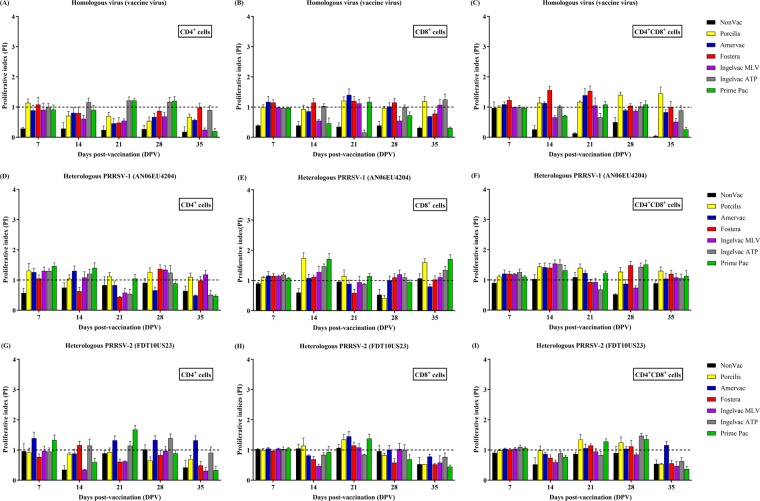
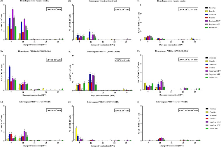
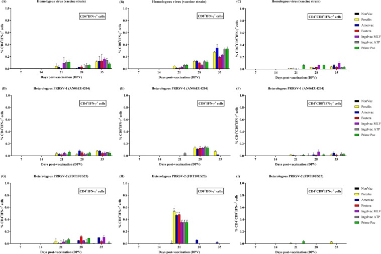
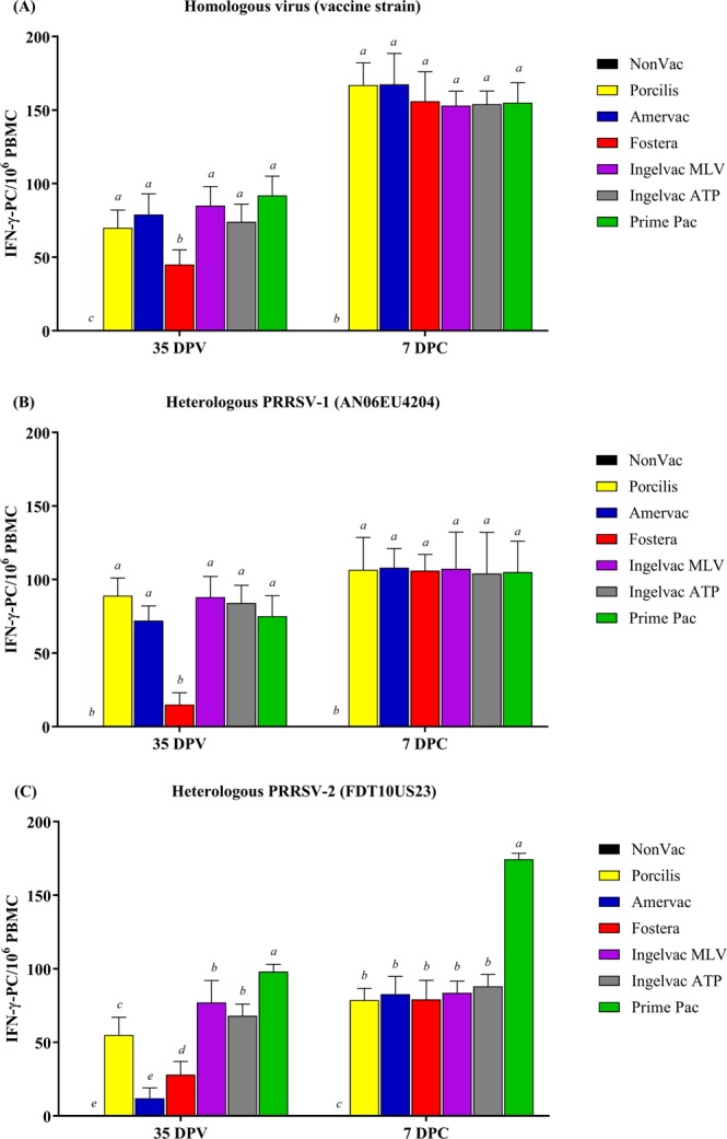
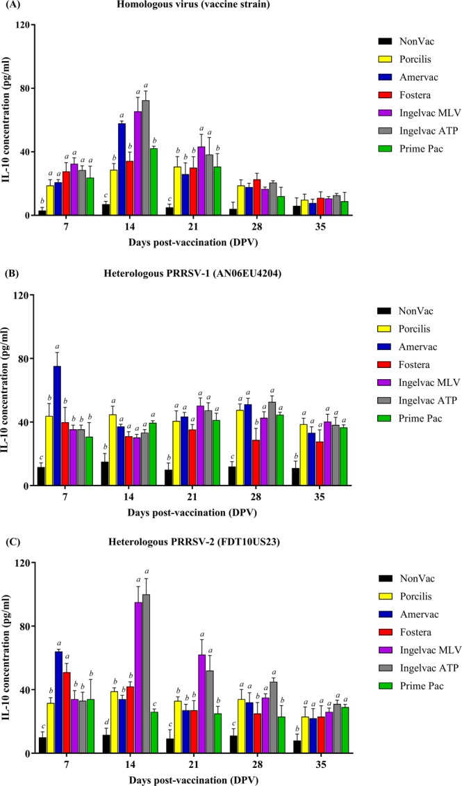
Cell-mediated immunity (CMI), IL-10, and the protective efficacy of modified-live porcine reproductive and respiratory syndrome virus (PRRSV) vaccines (MLV) against co-challenge with PRRSV-1 and PRRSV-2 (HP-PRRSV) were investigated. Seventy, PRRSV-free, 3-week old, pigs were allocated into 7 groups. Six groups were intramuscularly vaccinated with MLV, including Porcilis (PRRSV-1 MLV, MSD Animal Health, The Netherlands), Amervac (PRRSV-1 MLV, Laboratorios Hipra, Spain), Fostera (PRRSV-2 MLV, Zoetis, USA), Ingelvac PRRS MLV and Ingelvac PRRS ATP (PRRSV-2, Boehringer Ingelheim, USA), and Prime Pac PRRS (PRRSV-2 MLV, MSD Animal Health, The Netherlands). Unvaccinated pigs were left as control. Lymphocyte proliferative response, IL-10 and IFN-γ production were determined. At 35 days post-vaccination (DPV), all pigs were inoculated intranasally with 2 ml of each PRRSV-1 (105.4 TCID50/ml) and PRRSV-2 (105.2 TCID50/ml, HP-PRRSV). Following challenge, sera were quantitatively assayed for PRRSV RNA. Pigs were necropsied at 7 days post-challenge. Viremia, macro- and microscopic lung lesion together with PRRSV antigen presence were evaluated in lung tissues. The results demonstrated that, regardless of vaccine genotype, CMI induced by all MLVs was relatively slow. Increased production of IL-10 in all vaccinated groups was observed at 7 and 14 DPV. Pigs in Amervac, Ingelvac MLV and Ingelvac ATP groups had significantly higher levels of IL-10 compared to Porcilis, Fostera and Prime Pac groups at 7 and 14 DPV. Following challenge, regardless to vaccine genotype, vaccinated pigs had significantly lower lung lesion scores and PRRSV antigens than those in the control group. Both PRRSV-1 and PRRSV-2 RNA were significantly reduced. Prime Pac pigs had lowest PRRSV-1 and PRRSV-2 RNA in serum, and micro- and macroscopic lung lesion scores (p < 0.05) compared to other vaccinated groups. In conclusion, PRRSV MLVs, regardless of vaccine genotype, can reduce viremia and lung lesions following co-challenge with PRRSV-1 and PRRSV-2 (HP-PRRSV). The main difference between PRRSV MLV is the production of IL-10 following vaccination.


Conflict of interest statement

The authors declare no competing interests.

Figures

Figure 1
Figure 1
Lymphocyte proliferative index (PI) following vaccination. (AC) Homologous virus (vaccine strain), (DF) heterologous PRRSV-1 (AN06EU4204), and (GI) heterologous PRRSV-2 (FDT10US23), respectively. The lymphocyte populations were identified by flow cytometry using CFSE and cell surfaces staining, including CD4+ cells (A,D,G), CD8+ cells (B,E,H), and CD4+CD8+ cells (C,F,I), respectively. Values are expressed as mean±SEM. Dash lines indicate the cut-off level.
Figure 2
Figure 2
Lymphocyte populations producing IL-10 following vaccination. (AC) Homologous virus (vaccine strain), (DF) heterologous PRRSV-1 (AN06EU4204), and (GI) heterologous PRRSV-2 (FDT10US23), respectively. The lymphocyte populations producing IL-10 were identified by flow cytometry using cell surfaces and intracellular IL-10 staining, including CD4+IL-10+ cells (A,D,G), CD8+IL-10+ cells (B,E,H), and CD4+CD8+IL-10+ cells (C,F,I), respectively. Values are expressed as mean±SEM. Results were compared using two-way ANOVA multiple comparison test. Lowercase letters represent significant differences between treatment groups (p < 0.05) at each day post vaccination.
Figure 3
Figure 3
Lymphocyte populations producing IFN-γ following vaccination. (AC) homologous virus (vaccine strain), (DF) heterologous PRRSV-1 (AN06EU4204), and (GI) heterologous PRRSV-2 (FDT10US23), respectively. The lymphocyte populations producing IFN-γ were identified by flow cytometry using cell surfaces and intracellular IFN-γ staining, including CD4+IFN-γ+ cells (A,D,G), CD8+IFN-γ+ cells (B,E,H), and CD4+CD8+IFN-γ+ cells (C,F,I), respectively. Values are expressed as mean±SEM. Results were compared using two-way ANOVA multiple comparison test. Lowercase letters represent significant differences between treatment groups (p < 0.05) at each day post vaccination.
Figure 4
Figure 4
Evaluation of PRRSV-specific IFN-γ-PC following vaccination and at 7 days post-challenge (DPC) using in vitro stimulation. (A) Homologous virus (vaccine strain), (B) heterologous PRRSV-1 (AN06EU4204), and (C) heterologous PRRSV-2 (FDT10US23), respectively. Values are expressed as mean±SEM. Results were compared using two-way ANOVA multiple comparison test. Lowercase letters represent significant differences between treatment groups (p < 0.05) at each day post vaccination.
Figure 5
Figure 5
Quantification of porcine IL-10 in supernatant of stimulated PBMC following vaccination. (A) Homologous virus (vaccine strain), (B) heterologous PRRSV-1 (AN06EU4204), and (C) heterologous PRRSV-2 (FDT10US23), respectively. Values are expressed as mean ± SEM. Results were compared using two-way ANOVA multiple comparison test. Lowercase letters represent significant differences between treatment groups (p < 0.05) at each day post vaccination.

Similar articles

Cited by

References

    1. Cavanagh D. Nidovirales: a new order comprising Coronaviridae and Arteriviridae. Arch. Virol. 1997;142:629–633. - PubMed
    1. Nelson EA, et al. Differentiation of U.S. and European isolates of porcine reproductive and respiratory syndrome virus by monoclonal antibodies. J. Clin. Microbiol. 1993;31:3184–3189. doi: 10.1128/JCM.31.12.3184-3189.1993. - DOI - PMC - PubMed
    1. Stadejek T, et al. Definition of subtypes in the European genotype of porcine reproductive and respiratory syndrome virus: nucleocapsid characteristics and geographical distribution in Europe. Arch. Virol. 2008;153:1479–1488. doi: 10.1007/s00705-008-0146-2. - DOI - PubMed
    1. Shi M, et al. Phylogeny-based evolutionary, demographical, and geographical dissection of North American type 2 porcine reproductive and respiratory syndrome viruses. J. Virol. 2010;84:8700–8711. doi: 10.1128/JVI.02551-09. - DOI - PMC - PubMed
    1. Nilubol D, Tripipat T, Hoonsuwan T, Tipsombatboon P, Piriyapongsa J. Genetic diversity of the ORF5 gene of porcine reproductive and respiratory syndrome virus (PRRSV) genotypes I and II in Thailand. Arch. Virol. 2013;158:943–953. doi: 10.1007/s00705-012-1573-7. - DOI - PubMed

Publication types

MeSH terms