Multiple Complex Odontomas of the Mandible: A Rare Case Report and Literature Review
- PMID: 32015661
- PMCID: PMC6974989
- DOI: 10.4103/ccd.ccd_463_18
Multiple Complex Odontomas of the Mandible: A Rare Case Report and Literature Review
Abstract
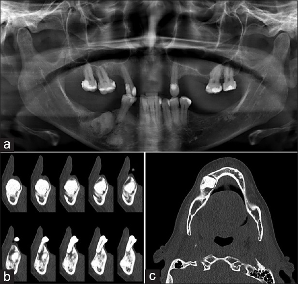
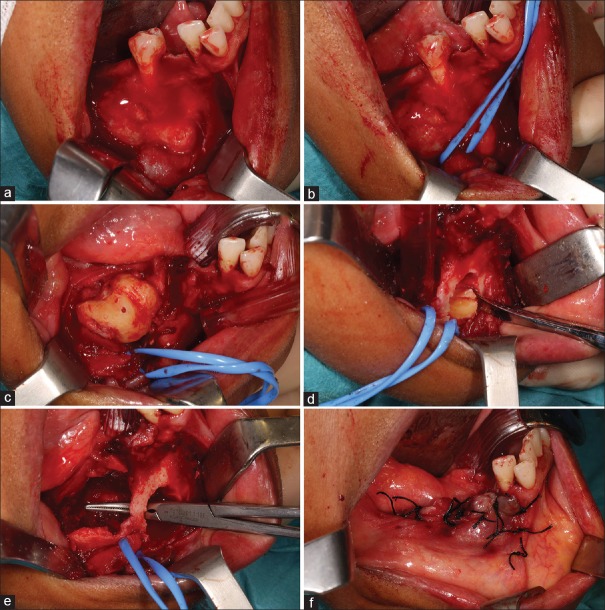
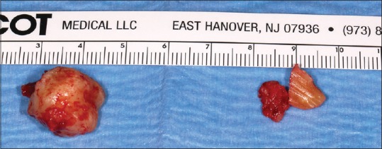
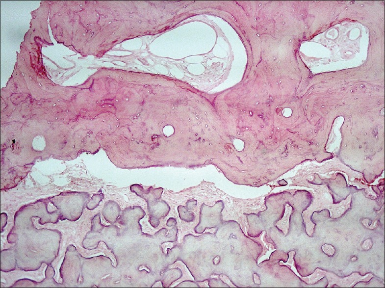
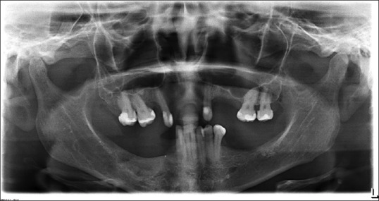
A 53-year-old female appeared with pain in the right mandible ramus, for the past 9 months, after tooth extraction. Clinical and radiological examination using conventional and advanced computerized tomography diagnostic imaging led to a provisional diagnosis of multiple complex odontomas. Complete conservative excision of the lesion was performed. The clinical diagnosis was confirmed histopathologically. Postoperative period was uneventful with no evidence of recurrence. According to an extensive literature review, this report describes the oldest patient ever diagnosed with multiple odontomas in the literature.
Keywords: Complex odontoma; hamartoma; mandible; multiple odontomas; odontogenic tumor.
Copyright: © 2019 Contemporary Clinical Dentistry.
Conflict of interest statement
There are no conflicts of interest.
Figures
Similar articles
-
Rare giant complex composite odontoma of mandible in mixed dentition: Case report with 3-year follow-up and literature review.Ann Med Surg (Lond). 2022 Feb 7;74:103355. doi: 10.1016/j.amsu.2022.103355. eCollection 2022 Feb. Ann Med Surg (Lond). 2022. PMID: 35198177 Free PMC article.
-
ODONTOMAS: PEDIATRIC CASE REPORT AND REVIEW OF THE LITERATURE.Acta Clin Croat. 2021 Mar;60(1):146-152. doi: 10.20471/acc.2021.60.01.22. Acta Clin Croat. 2021. PMID: 34588736 Free PMC article. Review.
-
Giant complex odontoma in the posterior mandible: A case report and literature review.Imaging Sci Dent. 2018 Dec;48(4):289-293. doi: 10.5624/isd.2018.48.4.289. Epub 2018 Dec 20. Imaging Sci Dent. 2018. PMID: 30607354 Free PMC article.
-
Complex Odontoma at an Unusual Site in a Child: A Case Report.Int J Clin Pediatr Dent. 2021 May-Jun;14(3):438-440. doi: 10.5005/jp-journals-10005-1950. Int J Clin Pediatr Dent. 2021. PMID: 34720522 Free PMC article.
-
Erupted odontomas: a report of three cases and review of the literature.Med Oral Patol Oral Cir Bucal. 2009 Jun 1;14(6):E299-303. Med Oral Patol Oral Cir Bucal. 2009. PMID: 19300370 Review.
Cited by
-
Rare giant complex composite odontoma of mandible in mixed dentition: Case report with 3-year follow-up and literature review.Ann Med Surg (Lond). 2022 Feb 7;74:103355. doi: 10.1016/j.amsu.2022.103355. eCollection 2022 Feb. Ann Med Surg (Lond). 2022. PMID: 35198177 Free PMC article.
-
ODONTOMAS: PEDIATRIC CASE REPORT AND REVIEW OF THE LITERATURE.Acta Clin Croat. 2021 Mar;60(1):146-152. doi: 10.20471/acc.2021.60.01.22. Acta Clin Croat. 2021. PMID: 34588736 Free PMC article. Review.
-
Management of Mandibular Compound Odontoma With Numbness in the Lower Jaw.Cureus. 2023 Dec 30;15(12):e51315. doi: 10.7759/cureus.51315. eCollection 2023 Dec. Cureus. 2023. PMID: 38288236 Free PMC article.
-
Ameloblastic fibro-odontoma or complex odontoma masquerading as gingival enlargement.J Indian Soc Periodontol. 2021 Sep-Oct;25(5):438-442. doi: 10.4103/jisp.jisp_778_20. Epub 2021 Aug 30. J Indian Soc Periodontol. 2021. PMID: 34667389 Free PMC article.
-
Sturge-Weber syndrome with cemento-ossifying fibroma in the maxilla and giant odontoma in the mandible: A case report.Heliyon. 2024 Apr 15;10(8):e29445. doi: 10.1016/j.heliyon.2024.e29445. eCollection 2024 Apr 30. Heliyon. 2024. PMID: 38660248 Free PMC article.
References
-
- Regezi J, Sciubba J. 5th ed. St. Louis: Saunder; 2007. Oral Pathology: Clinical Pathologic Correlations.
-
- Barnes L, Eveson JW, Reichart P, Sidransky D. WHO Press; 2005. Pathology and Genetics of Head and Neck Tumours: World Health Organization Classification Tumour: 163.
-
- Buchner A, Merrell PW, Carpenter WM. Relative frequency of central odontogenic tumors: A study of 1,088 cases from Northern California and comparison to studies from other parts of the world. J Oral Maxillofac Surg. 2006;64:1343. - PubMed
-
- Jing W, Xuan M, Lin Y, Wu L, Liu L, Zheng X, et al. Odontogenic tumours: A retrospective study of 1642 cases in a Chinese population. Int J Oral Maxillofac Surg. 2007;36:20–5. - PubMed
-
- Luo HY, Li TJ. Odontogenic tumors: A study of 1309 cases in a Chinese population. Oral Oncol. 2009;45:706–11. - PubMed
Publication types
LinkOut - more resources
Full Text Sources
Miscellaneous