Solitary Bone Cyst of Posterior Maxilla: A Rare Presentation
- PMID: 32015662
- PMCID: PMC6975003
- DOI: 10.4103/ccd.ccd_470_18
Solitary Bone Cyst of Posterior Maxilla: A Rare Presentation
Abstract
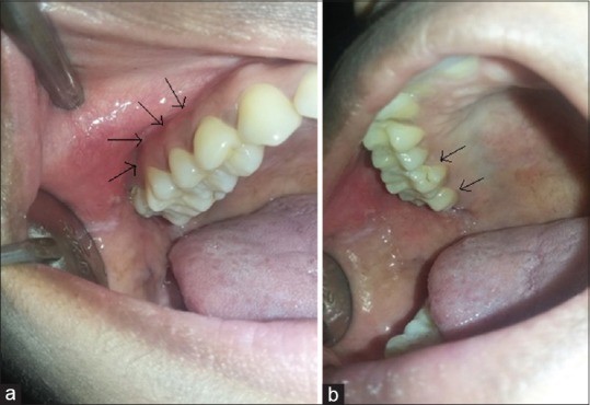
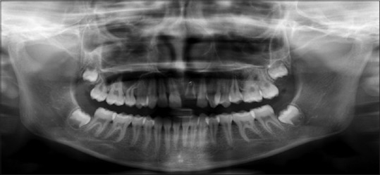
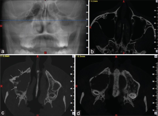
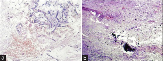
Solitary bone cyst (SBC) is an uncommon, nonneoplastic osseous lesion that mainly affects metaphysis of long bones and rarely presents in jaws. Due to the lack of true epithelial lining, it is considered as a pseudocyst. It is generally asymptomatic and often discovered incidentally during routine radiographic examination as well-defined unilocular or multilocular radiolucent lesion in the posterior mandible mainly in the first two decades of life. Here, we report a very rare case of a 15-year-old female patient having a lesion in the posterior maxilla with clinical, radiological, and histopathological presentations of SBC.
Keywords: Maxilla; solitary bone cyst; unilocular radiolucency.
Copyright: © 2019 Contemporary Clinical Dentistry.
Conflict of interest statement
There are no conflicts of interest.
Figures
Similar articles
-
[Simple bone cyst in the mandible--a rare occurrence in an elderly patient].Refuat Hapeh Vehashinayim (1993). 2006 Jan;23(1):27-30, 69. Refuat Hapeh Vehashinayim (1993). 2006. PMID: 16599330 Hebrew.
-
Traumatic bone cyst of mandible.J Maxillofac Oral Surg. 2015 Jun;14(2):466-9. doi: 10.1007/s12663-010-0114-8. Epub 2010 Nov 25. J Maxillofac Oral Surg. 2015. PMID: 26028875 Free PMC article.
-
Traumatic Bone Cyst of Mandible: A Case Report of Rare Entity and Review of Literature.Contemp Clin Dent. 2019 Jan-Mar;10(1):3-8. doi: 10.4103/ccd.ccd_489_18. Contemp Clin Dent. 2019. PMID: 32015634 Free PMC article. Review.
-
Traumatic bone cyst of mandible: a case series.J Med Case Rep. 2019 Sep 18;13(1):300. doi: 10.1186/s13256-019-2220-7. J Med Case Rep. 2019. PMID: 31530284 Free PMC article. Review.
-
Solitary bone cysts-A rare occurrence with bilaterally symmetrical presentation.J Oral Maxillofac Pathol. 2014 Sep-Dec;18(3):481. doi: 10.4103/0973-029X.151366. J Oral Maxillofac Pathol. 2014. PMID: 25949013 Free PMC article.
Cited by
-
Solitary bone cyst in a 57-year-old woman - A case report.Int J Surg Case Rep. 2025 Jul 26;134:111736. doi: 10.1016/j.ijscr.2025.111736. Online ahead of print. Int J Surg Case Rep. 2025. PMID: 40729805 Free PMC article.
References
-
- Harnet JC, Lombardi T, Klewansky P, Rieger J, Tempe MH, Clavert JM, et al. Solitary bone cyst of the jaws: A review of the etiopathogenic hypotheses. J Oral Maxillofac Surg. 2008;66:2345–8. - PubMed
-
- Titsinides S, Kalyvas D. Traumatic bone cyst of the jaw: A case report and review of previous studies. J Dent Health Oral Disord Ther. 2016;5:00167.
-
- Shear M. 3rd ed. Oxford: Wright PSG; 1992. Cysts of the Oral Regions.
-
- Cortell-Ballester I, Figueiredo R, Berini-Aytés L, Gay-Escoda C. Traumatic bone cyst: A retrospective study of 21 cases. Med Oral Patol Oral Cir Bucal. 2009;14:E239–43. - PubMed