

Biomechanical and Functional Improvements Gained by Proximal Tibia Osteotomy Correction of Genu Varum in Patients with Knee Pain
- PMID: 32015738
- PMCID: PMC6973828
- DOI: 10.1007/s11420-019-09670-6
Biomechanical and Functional Improvements Gained by Proximal Tibia Osteotomy Correction of Genu Varum in Patients with Knee Pain
Abstract
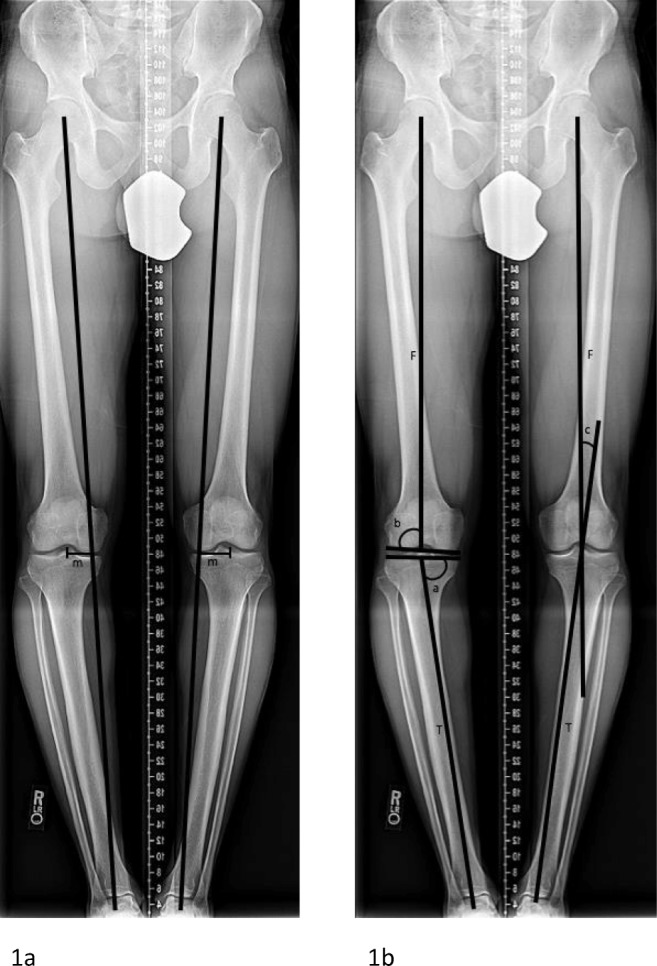
Background: Mechanical axis malalignment contributes to abnormal forces across the knee joint. Genu varum, or increased medial mechanical axis deviation (MAD), increases force transmission and contact pressures to the medial compartment. With increasing MAD and femoral-tibial mechanical axis angle (MAA), contact forces within the medial or lateral compartment of the knee significantly increase with increasing deformity. This may lead to knee pain, further deformity, and medial compartment degenerative joint disease, which can interfere with participation in sports and diminish quality of life.
Purposes/questions: We sought to evaluate patients with knee pain with bilateral genu varum and determine the effect of bilateral proximal tibial osteotomies on knee biomechanics, deformity correction, and functional outcomes.
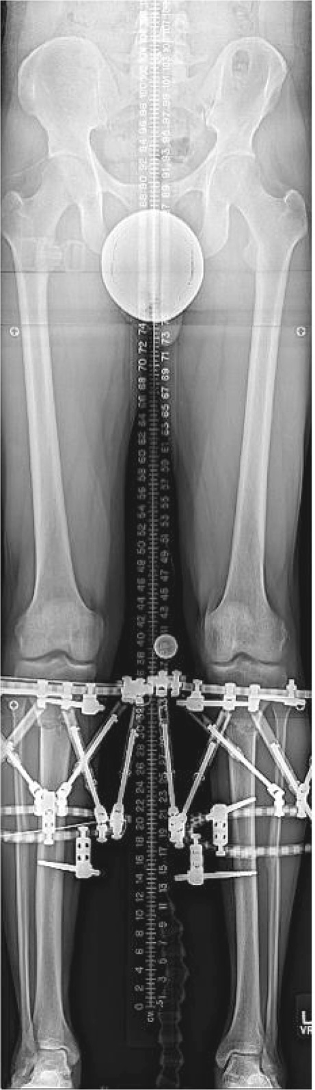
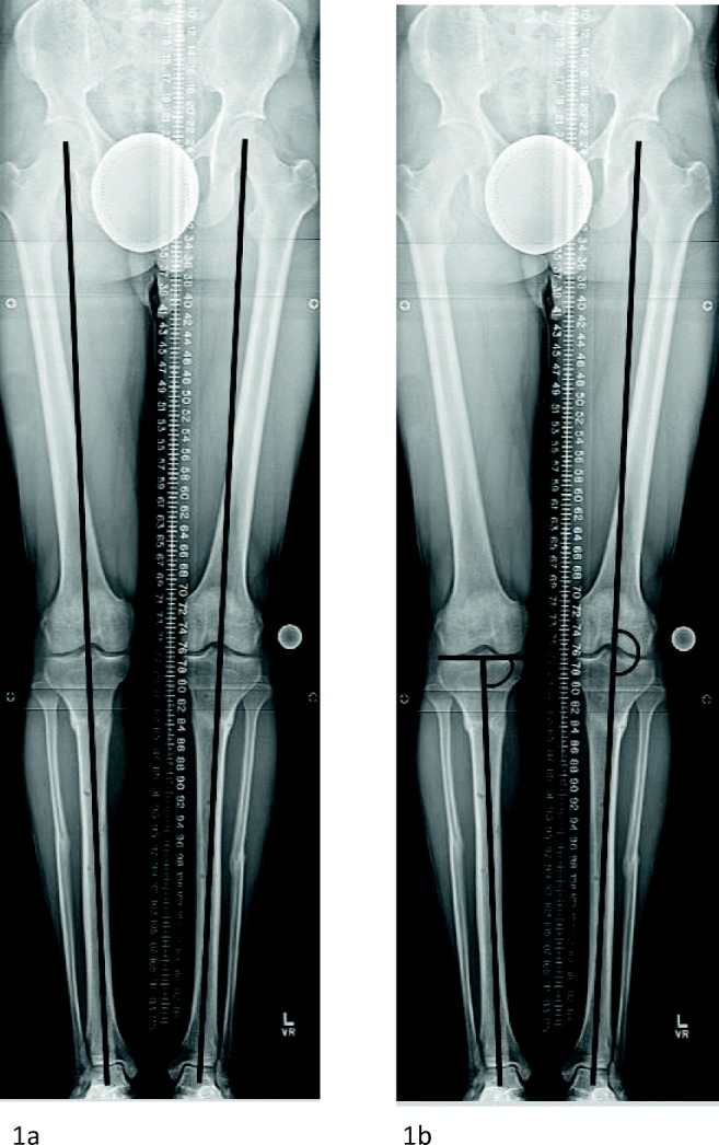
Methods: This was a single-center, prospective study of eight limbs in four patients. Consecutive patients presenting with knee pain and bilateral genu varum originating from the proximal tibia were included. All patients underwent staged bilateral proximal tibial osteotomies with gradual deformity correction with an external fixator. Subjects underwent a three-dimensional (3D) instrumented motion analysis during level walking. A 3D lower extremity model was built and bilateral knee frontal plane kinematics and kinetics during the stance phase of gait were determined. Radiographic analysis was performed including assessment of MAD, MAA, and medial proximal tibial angle (MPTA). Functional outcomes were assessed with the Knee Injury and Osteoarthritis Outcome Score (KOOS), the 36-item Short-Form Survey (SF-36), the Lower Limb Questionnaire (LLQ), and a visual analog scale (VAS) for pain.
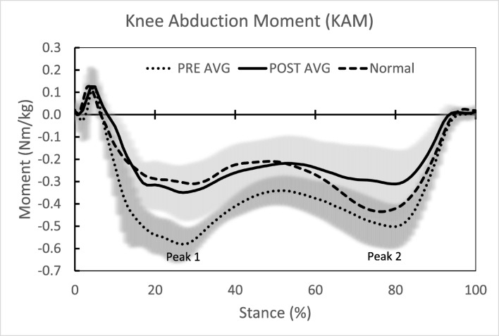
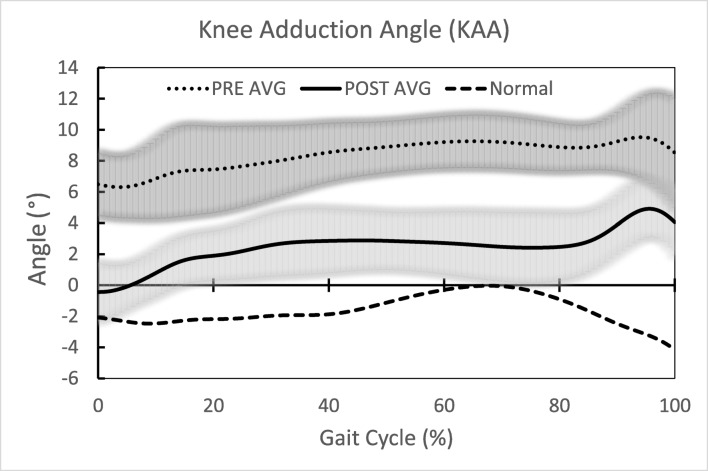
Results: The average time in the external fixator for a single limb was 97 days. The average follow-up period was 310 days. All biomechanical outcomes significantly improved, including knee adduction angle (7.8° to 1.8°), knee adduction moments (first peak, - 0.450 to - 0.281 nm/kg, and second peak, - 0.381 to - 0.244 nm/kg), and knee adduction moment impulse (- 0.233 to - 0.150 nm s/kg). There was a significant improvement in MAA, MAD, and MPTA. All patients showed qualitative improvement in mean scores on VAS (11.8 to 0.0), LLQ (77 to 93), KOOS (64 to 84), and SF-36 (71 to 88).
Conclusion: These findings suggest that bilateral proximal tibial osteotomy may be effective in improving knee biomechanics during gait and correcting mechanical alignment in patients with bilateral genu varum. Patients also demonstrated improvement in functional outcome scores. This technique should thus be considered in patients with varus knee osteoarthritis in the setting of genu varum to alleviate symptoms and potentially decrease further clinical deterioration.
Keywords: external fixation; gait biomechanics; knee pain; osteoarthritis; proximal tibial osteotomy; varus.
© The Author(s) 2019.
Conflict of interest statement
Conflict of InterestRachael J. Da Cunha, MD, FRCSC, Andrew P. Kraszewski, PhD, and Howard J. Hillstrom, PhD, declare that they have no conflicts of interest. Austin T. Fragomen, MD, reports being a paid consultant to Smith & Nephew, Synthes, Globus, and NuVasive, outside the submitted work. S. Robert Rozbruch, MD, reports being a paid consultant to Smith & Nephew, Stryker, and NuVasive, outside the submitted work.
Figures
Similar articles
-
Medial Closing Wedge High Tibial Osteotomy Accurately Corrects Genu Valgum without Iatrogenic Deformity or Complications: A Consecutive Series of Thirty-one Procedures.Strategies Trauma Limb Reconstr. 2024 May-Aug;19(2):82-86. doi: 10.5005/jp-journals-10080-1620. Epub 2024 Aug 14. Strategies Trauma Limb Reconstr. 2024. PMID: 39359363 Free PMC article.
-
Does the Surgical Correction of Tibial Torsion with Genu Varum Produce Outcomes Similar to Those in Varus Correction Alone?J Knee Surg. 2018 Apr;31(4):359-369. doi: 10.1055/s-0037-1603797. Epub 2017 Jun 24. J Knee Surg. 2018. PMID: 28646823
-
High Tibial Osteotomy for Genu Varum in Adults: Do Proprietary Implants Limit the Quality of Correction?Strategies Trauma Limb Reconstr. 2020 Jan-Apr;15(1):13-22. doi: 10.5005/jp-journals-10080-1449. Strategies Trauma Limb Reconstr. 2020. PMID: 33363636 Free PMC article.
-
High tibial osteotomy: evolution of research and clinical applications--a Canadian experience.Knee Surg Sports Traumatol Arthrosc. 2013 Jan;21(1):23-31. doi: 10.1007/s00167-012-2218-9. Epub 2012 Sep 28. Knee Surg Sports Traumatol Arthrosc. 2013. PMID: 23052112 Review.
-
Biomechanics of high tibial osteotomy.Knee Surg Sports Traumatol Arthrosc. 2013 Jan;21(1):197-205. doi: 10.1007/s00167-012-2122-3. Epub 2012 Jul 7. Knee Surg Sports Traumatol Arthrosc. 2013. PMID: 22773067 Review.
Cited by
-
Correlation of lower limb alignment with medial mensical extrusion in knee osteoarthritis.Arch Orthop Trauma Surg. 2024 Nov;144(11):4819-4826. doi: 10.1007/s00402-024-05568-z. Epub 2024 Sep 21. Arch Orthop Trauma Surg. 2024. PMID: 39305324
-
The Relationships between Coronal Plane Alignments and Patient-Reported Outcomes Following High Tibial Osteotomy: A Systematic Review.Cartilage. 2021 Dec;13(1_suppl):132S-146S. doi: 10.1177/19476035211007903. Epub 2021 Apr 22. Cartilage. 2021. PMID: 33884908 Free PMC article.
-
Gradual Correction of Valgus Deformities of the Tibia Using a Monolateral External Fixator.Strategies Trauma Limb Reconstr. 2023 May-Aug;18(2):123-132. doi: 10.5005/jp-journals-10080-1585. Strategies Trauma Limb Reconstr. 2023. PMID: 37942429 Free PMC article.
-
Opening wedge high tibial osteotomy for medial compartment knee osteoarthritis: Planning and improving outcomes: Case series and literature review.J Clin Orthop Trauma. 2022 Dec 5;36:102085. doi: 10.1016/j.jcot.2022.102085. eCollection 2023 Jan. J Clin Orthop Trauma. 2022. PMID: 36654729 Free PMC article.
-
Gait analysis and knee joint kinematics before a and 6 month after of corrective valgus osteotomy at patients with medial knee arthritis.Int Orthop. 2022 Jul;46(7):1573-1582. doi: 10.1007/s00264-022-05370-9. Epub 2022 Apr 13. Int Orthop. 2022. PMID: 35416482
References
-
- Adili A, Bhandari M, Giffin R, Whately C, Kwok DC. Valgus high tibial osteotomy. Comparison between an Ilizarov and a Coventry wedge technique for the treatment of medial compartment osteoarthritis of the knee. Knee Surg Sports Traumatol Arthrosc. 2002;10(3):169–176. doi: 10.1007/s00167-001-0250-2. - DOI - PubMed
-
- Coventry MB. Upper tibial osteotomy. Clin Orthop Relat Res. 1984;182:46–52. - PubMed
LinkOut - more resources
Full Text Sources
