

Dataset on interactions of membrane active agents with lipid bilayers
- PMID: 32016146
- PMCID: PMC6992954
- DOI: 10.1016/j.dib.2020.105138
Dataset on interactions of membrane active agents with lipid bilayers
Abstract
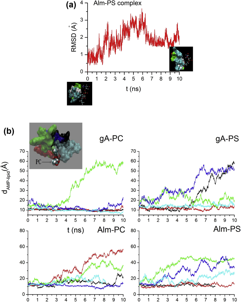
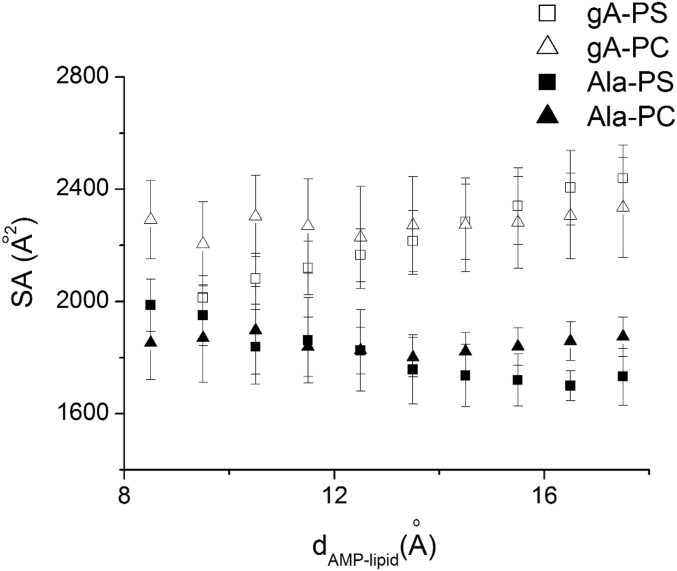
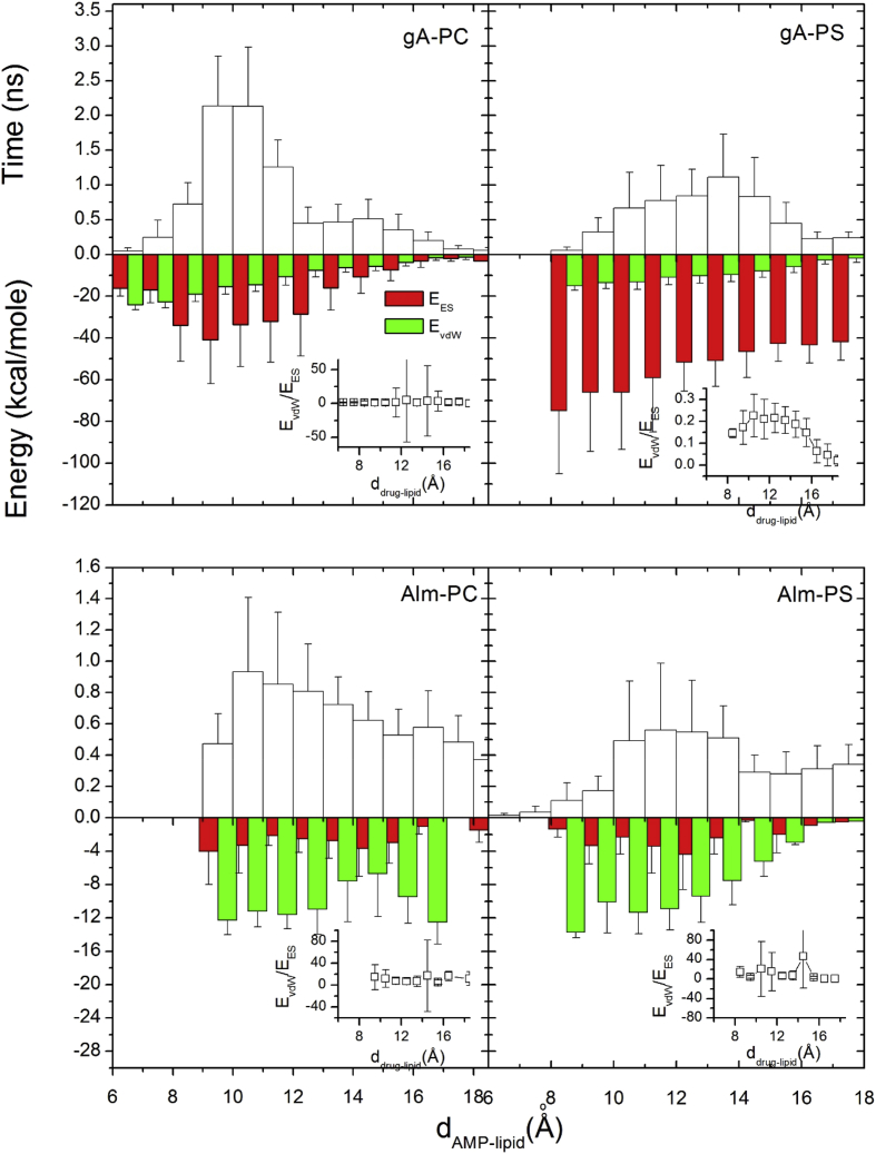
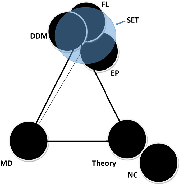
We address drug interactions with lipids using in silico simulations and in vitro experiments. The data article provides extended explanations on molecular mechanisms behind membrane action of membrane-active agents (MAAs): antimicrobial peptides and chemotherapy drugs. Complete interpretation of the data is found in the associated original article 'charge-based interactions of antimicrobial peptides and general drugs with lipid bilayers' [1]. Data on molecular dynamic simulations of the drug lipid complexes are provided. Additional data and information are provided here to explain the connectivity among various information and techniques used for understanding of the membrane action and/or binding of MAAs including aptamers. Brief explanation has been provided on the possibility of achieving a converted triangle from newly discovered quadrangle, sides of which explain four different phenomena: 'membrane effects', 'detection and quantification', 'origin of energetics' and 'structure stability' while drug effects occur. Triangle or quadrangle corners represent various techniques that were applied.
Keywords: Direct detection method; Drugs; Electrostatic interactions; Ion channel; Lipid membrane.
© 2020 The Author(s).
Figures
References
-
- Ashrafuzzaman M., Tseng C.Y., Tuszynski J. Charge-based interactions of antimicrobial peptides and general drugs with lipid bilayers. J. Mol. Graph. Model. 2020;95 - PubMed
-
- Ashrafuzzaman M., Tseng C.Y., Tuszynski J. Regulation of channel function due to physical energetic coupling with a lipid bilayer. Biochem. Biophys. Res. Comn. 2014;445:463–468. - PubMed
-
- Tseng C.Y., Ashrafuzzaman M., Mane J., Kapty J., Mercer J., Tuszynski J.A. Entropic fragment based approach to aptamer design. Chem. Biol. Drug Des. 2011;78:1–13. - PubMed
-
- Ashrafuzzaman M., Tseng C.Y., Duszyk M., Tuszynski J. Chemotherapy drugs form ion pores in membranes due to physical interactions with lipids. Chem. Biol. Drug Des. 2012;80:992–1002. - PubMed
LinkOut - more resources
Full Text Sources
