. 2020 Feb 3;6(1):34.
doi: 10.1186/s40792-020-0784-5.

Metachronous rupture of a residual pancreaticoduodenal aneurysm after release of the median arcuate ligament: a case report

Affiliations

Metachronous rupture of a residual pancreaticoduodenal aneurysm after release of the median arcuate ligament: a case report

Nana Kimura et al. Surg Case Rep. .

Abstract

Background: Multiple pancreaticoduodenal artery aneurysms in association with median arcuate ligament syndrome (MALS) are relatively rare. A treatment option, such as a median arcuate ligament (MAL) release or embolization of the aneurysms, should be considered in such cases, but the treatment criteria remain unclear.

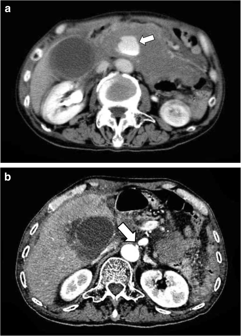
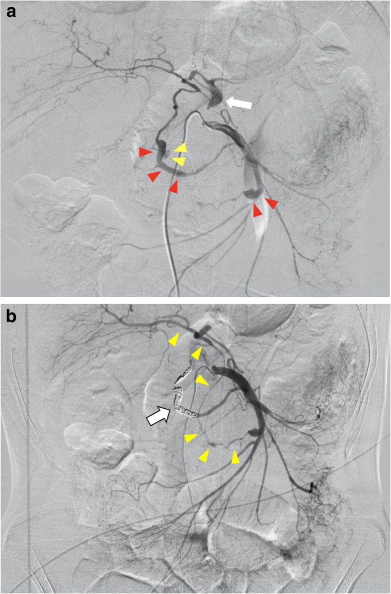
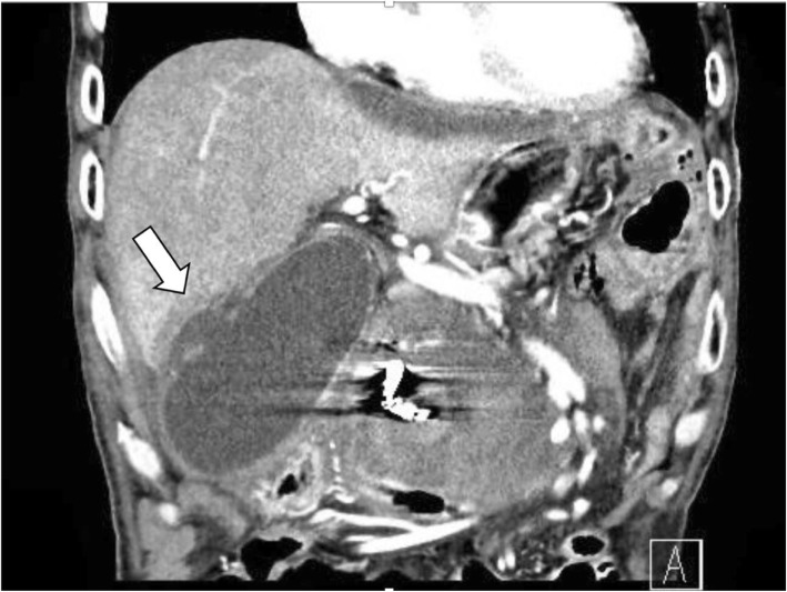
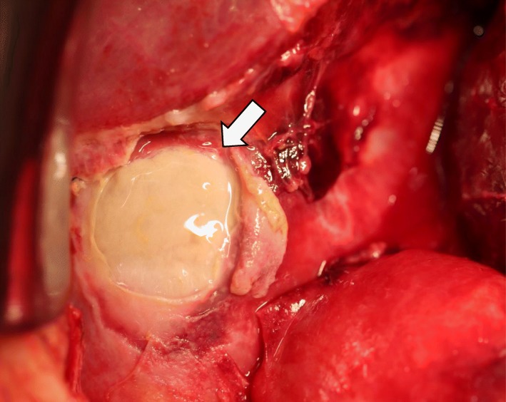
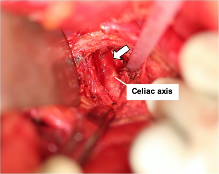
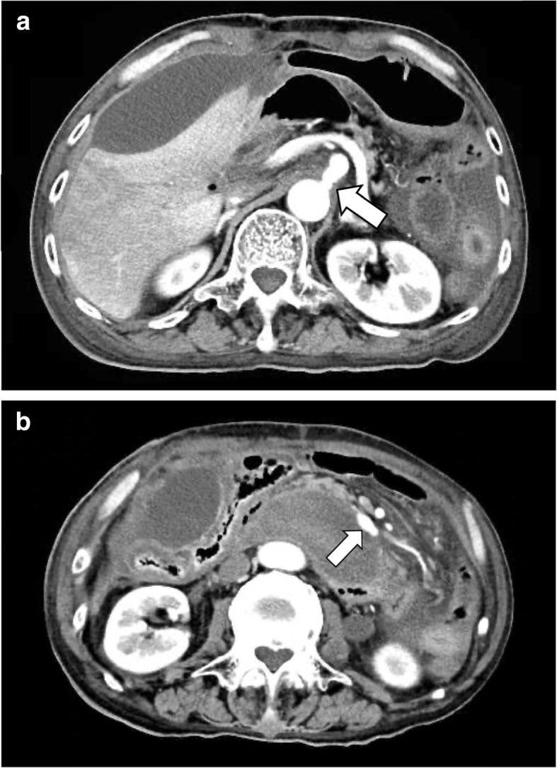
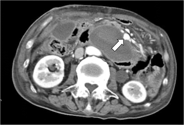
Case report: A 75-year-old man was transferred to our hospital because of a ruptured pancreaticoduodenal aneurysm. Emergency angiography showed stenosis of the root of the celiac axis (CA), a ruptured aneurysm of the posterior inferior pancreaticoduodenal artery (PIPDA), and an unruptured aneurysm of the anterior inferior pancreaticoduodenal artery (AIPDA). Coil embolization of the PIPDA was performed. Five days after embolization, the gallbladder became necrotic due to decreased blood flow in the CA region, and an emergency operation was performed. We performed a cholecystectomy and released the MAL to normalize the blood flow of the CA region. However, the patient died on postoperative day 8 because of rupture of the untreated aneurysm of the AIPDA.

Conclusions: This is the first report of metachronous ruptures of multiple pancreaticoduodenal aneurysms due to MALS, even after a MAL release. Although rare, a residual aneurysm in the pancreatic head region may need to be embolized quickly.

Keywords: Median arcuate ligament syndrome; Metachronous rupture; Multiple aneurysm; Segmental arterial mediolysis.


Conflict of interest statement

The authors declare that they have no competing interests.

Figures

Fig. 1
Fig. 1
a Computed tomography taken at visiting a physician showed a hematoma, and inferior pancreaticoduodenal artery aneurysm rupture was suspected (shown by arrow). b Computed tomography taken at visiting a physician showed stenosis of the root of the celiac axis (shown by arrow)
Fig. 2
Fig. 2
a Emergency angiography revealed stenosis of the origin of the celiac artery (shown by arrow), and spindle-shaped dilatation and pseudoaneurysm formation were observed in posterior inferior pancreaticoduodenal artery to posterior superior pancreaticoduodenal artery and anterior inferior pancreaticoduodenal artery (shown by red arrowheads). Contrast medium extravasation from posterior inferior pancreaticoduodenal artery was observed (shown by yellow arrowheads). Irregular vasodilation and stenosis were observed in multiple arteries, which were considered to be the effect of segmental arterial mediolysis. b Embolization was performed on posterior inferior pancreaticoduodenal artery to posterior superior pancreaticoduodenal artery (shown by arrow). After coil embolization of the posterior inferior pancreaticoduodenal arterial aneurysm, the celiac arterial region was visualized from the anterior inferior pancreaticoduodenal artery via the gastroduodenal artery (shown by arrowheads)
Fig. 3
Fig. 3
Post-embolization computed tomography shows swollen gallbladder and encapsulated fluid retention around it (shown by arrow). It suggested that the wall was broken at the bottom of the gallbladder
Fig. 4
Fig. 4
The intraoperative finding showed the wall of the gallbladder was partially necrotic (shown by arrow)
Fig. 5
Fig. 5
The intraoperative identification of the median arcuate ligament (shown by arrow)
Fig. 6
Fig. 6
a The abdominal computed tomography on postoperative day 4 determined that the arterial diameter of the celiac axis has expanded slightly (shown by arrow). b The abdominal computed tomography on postoperative day 4 determined that there was no significant change in the size (13 × 7 mm) of the untreated aneurysm of the anterior inferior pancreaticoduodenal artery (shown by arrow)
Fig. 7
Fig. 7
Computed tomography after first vomiting showed extravasations from the untreated anterior inferior pancreaticoduodenal artery aneurysm (shown by arrowheads). An untreated anterior inferior pancreaticoduodenal artery aneurysm appeared to be rounded (13 × 11 mm)

References

    1. Kim EN, Lamb K, Relles D, Moudgill D, DiMuzio PJ, Eisenberg JA. Median arcuate ligament syndrome—review of this rare disease. JAMA Surg. 2016;151(5):471–477. doi: 10.1001/jamasurg.2016.0002. - DOI - PubMed
    1. Tracci MC. Median arcuate ligament compression of the mesenteric vasculature. Tech Vasc Interv Radiol. 2015;18(1):43–50. doi: 10.1053/j.tvir.2014.12.007. - DOI - PubMed
    1. Stanley J, Wakefield T, Graham L, Whitehouse W, Zelenock G, Lindenauer S. Clinical importance and management of splanchnic artery aneurysms. J Vasc Surg. 1986;3(5):836–840. doi: 10.1016/0741-5214(86)90059-5. - DOI - PubMed
    1. Armstrong MB, Stadtlander KS, Grove MK. Pancreaticoduodenal artery aneurysm associated with median arcuate ligament syndrome. Ann Vasc Surg. 2014;28(3):741.e1–741.e5. doi: 10.1016/j.avsg.2013.06.030. - DOI - PubMed
    1. Chivot C, Rebibo L, Robert B, Regimbeau JM, Yzet T. Ruptured pancreaticoduodenal artery aneurysms associated with celiac stenosis caused by the median arcuate ligament: a poorly known etiology of acute abdominal pain. Eur J Vasc Endovasc Surg. 2016;51(2):295–301. doi: 10.1016/j.ejvs.2015.10.025. - DOI - PubMed