

Cloning and functional testing of rhesus macaque (Macaca mulatta) IL-9 and IL-33
- PMID: 32017131
- PMCID: PMC7198344
- DOI: 10.1111/jmp.12464
Cloning and functional testing of rhesus macaque (Macaca mulatta) IL-9 and IL-33
Abstract
Background: IL-9 and IL-33 can profoundly influence immune responses. As a necessary first step toward defining their impact in the rhesus macaque model, we confirmed their endogenous expression and sequence identity and generated expression vectors for the recombinant expression of rhesus IL-9 and IL-33.
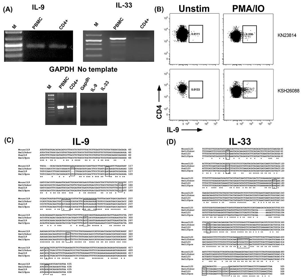
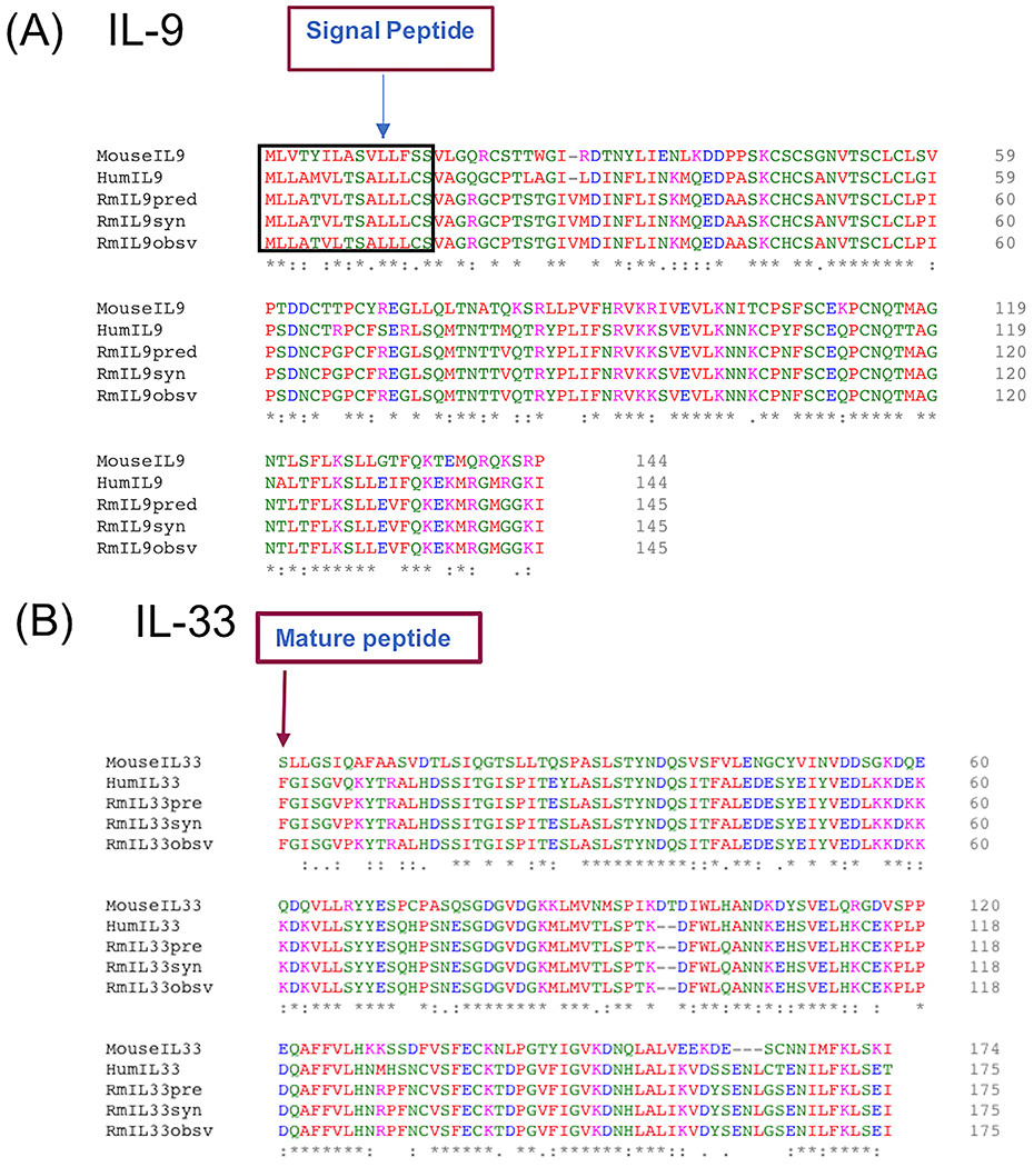
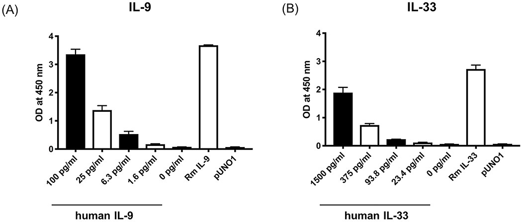
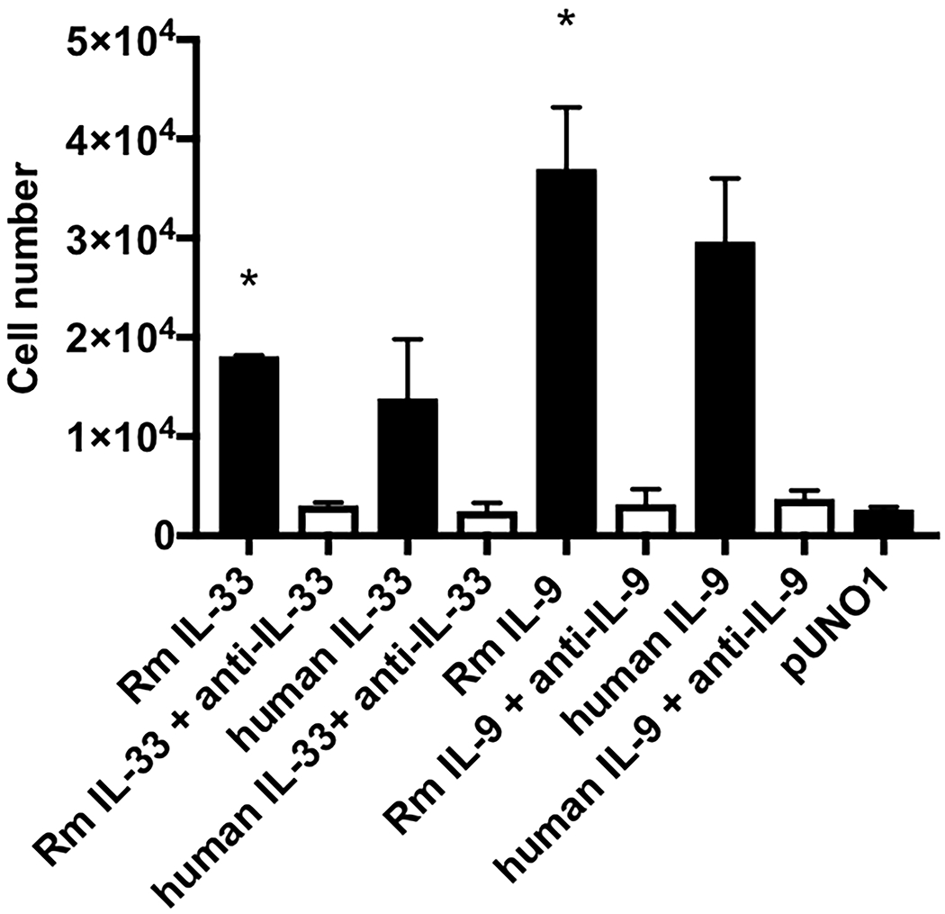
Methods: RT-PCR and Sanger sequencing was used to define the expression and sequences for rhesus IL-9 and IL-33. The resulting recombinant cytokines were tested by ELISA and proliferation assays.
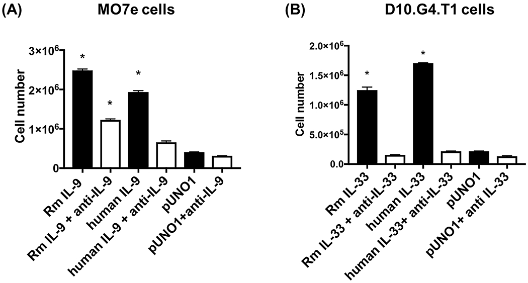
Results: Full-length rhesus IL-9 and the mature form of rhesus IL-33 share 78% and 73% nucleotide similarity, respectively, with humans. Both cytokines are expressed in lymphocytes, with IL-9 expression also evident in CD4+ T cells. Recombinantly expressed rhesus IL-9 and IL-33 were each biologically active in vitro, including enhancing the proliferation of a rhesus B cell line.
Conclusions: The recombinant rhesus IL-9 and IL-33 constructs produce biologically active cytokines that can act upon rhesus B cells.
Keywords: B cell; IL-33; IL-9; proliferation; rhesus.
© 2020 John Wiley & Sons A/S. Published by John Wiley & Sons Ltd.
Figures
References
-
- Hultner L, Druez C, Moeller J, Uyttenhove C, Schmitt E, Rude E, Dormer P, Van Snick J: Mast cell growth-enhancing activity (MEA) is structurally related and functionally identical to the novel mouse T cell growth factor P40/TCGFIII (interleukin 9). Eur J Immunol 1990; 20:1413–1416. - PubMed
-
- Gessner A, Blum H, Rollinghoff M: Differential regulation of IL-9-expression after infection with Leishmania major in susceptible and resistant mice. Immunobiology 1993; 189:419–435. - PubMed
-
- Dardalhon V, Awasthi A, Kwon H, Galileos G, Gao W, Sobel RA, Mitsdoerffer M, Strom TB, Elyaman W, Ho IC, Khoury S, Oukka M, Kuchroo VK: IL-4 inhibits TGF-beta-induced Foxp3+ T cells and, together with TGF-beta, generates IL-9+ IL-10+ Foxp3(−) effector T cells. Nat Immunol 2008; 9:1347–1355. - PMC - PubMed
Publication types
MeSH terms
Substances
Associated data
- Actions
- Actions
- Actions
- Actions
Grants and funding
LinkOut - more resources
Full Text Sources
Research Materials
