. 2020 Sep;84(3):1173-1183.
doi: 10.1002/mrm.28187. Epub 2020 Feb 4.

Non-contrast-enhanced abdominal MRA at 3 T using velocity-selective pulse trains

Affiliations

Non-contrast-enhanced abdominal MRA at 3 T using velocity-selective pulse trains

Dan Zhu et al. Magn Reson Med. 2020 Sep.

Abstract

Purpose: Most existing non-contrast-enhanced methods for abdominal MR arteriography rely on a spatially selective inversion (SSI) pulse with a delay to null both static tissue and venous blood, and are limited to small spatial coverage due to the sensitivity to slow arterial inflow. Velocity-selective inversion (VSI) based approach has been shown to preserve the arterial blood inside the imaging volume at 1.5 T. Recently, velocity-selective saturation (VSS) pulse trains were applied to suppress the static tissue and have been combined with SSI pulses for cerebral MR arteriography at 3 T. The aim of this study is to construct an abdominal MRA protocol with large spatial coverage at 3 T using advanced velocity-selective pulse trains.

Methods: Multiple velocity-selective MRA protocols with different sequence modules and 3D acquisition methods were evaluated. Sequences using VSS only as well as SSI+VSS and VSI+VSS preparations were then compared among a group of healthy young and middle-aged volunteers. Using MRA without any preparations as reference, relative signal ratios and relative contrast ratios of different vascular segments were quantitatively analyzed.

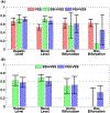
Results: Both SSI+VSS and VSI+VSS arteriograms achieved high artery-to-tissue and artery-to-vein relative contrast ratios above aortic bifurcation. The SSI+VSS sequence yielded lower signal at the bilateral iliac arteries than VSI+VSS, reflecting the benefit of the VSI preparation for imaging the distal branches.

Conclusion: The feasibility of noncontrast 3D MR abdominal arteriography was demonstrated on healthy volunteers using a combination of VSS pulse trains and SSI or VSI pulse.

Keywords: abdominal MRA; arteriography; non-contrast-enhanced MRA; velocity-selective pulse train.


Figures

FIGURE 1
FIGURE 1
A, Diagram of the abdominal velocity-selective (VS)-MRA sequences. A Fourier transform–based velocity-selective saturation (VSS) pulse train was placed right before fat suppression and 3D acquisition modules to suppress static tissue. Spatially selective inversion (SSI) or velocity-selective inversion (VSI) pulse with an inversion delay (dashed box) was applied to null venous blood signal for arteriography. Respiratory triggering was used before preparations. Diagram of the VSS pulse train (B) and its corresponding VS profile (C). Note that the longitudinal magnetization (Mz) for tissue (v = 0 cm/s) should be close to 0 when taking into account the T2 effect. Diagram of the VSI pulse train (D) and its corresponding VS profile (E)
FIGURE 2
FIGURE 2
Positioning of the MRA sequences used in this study. The yellow boxes indicate the imaging volumes for the different abdominal VS-MRA protocols with VSS, SSI+VSS, or VSI+VSS preparations, all with a 300-mm spatial coverage in the foot–head (FH) direction (A), and the vendor-provided SSI-based renal MRA protocol (B-TRANCE), with a 100-mm coverage in the FH direction (B). The blue boxes indicate the inversion bands applied in the SSI-VSS-prepared abdominal MRA scans (A) and the SSI-prepared renal MRA scans (B), both with default inversion delays of 1200 ms. The white boxes in (B) indicate the saturation bands applied for venous or fat suppression right before the acquisition
FIGURE 3
FIGURE 3
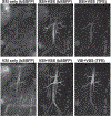
Coronal maximum intensity projection (MIP) arteriography from a 25-year-old female using SSI (top row) and VSI (bottom row) preparations. Comparisons were made among sequences applying SSI or VSI only with balanced SSFP (bSSFP) acquisition (left column), a combination of SSI or VSI with VSS using bSSFP (middle column), and turbo field-echo (TFE) (right column) acquisitions, respectively
FIGURE 4
FIGURE 4
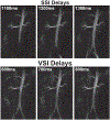
Coronal MIP arteriography from a 24-year-old male using SSI+VSS (top row) and VSI+VSS (bottom row) preparations with various inversion delays
FIGURE 5
FIGURE 5
Coronal MIP images of the reference scans without applying any of the VSS, SSI, or VSI preparation pulses (first column), full angiograms of both arteries and veins using the VSS pulse train for static tissue suppression (second column), and arteriograms using the SSI+VSS sequence (third column) and VSI+VSS sequence (fourth column), from a 25-year-old female (case 1, top row), a 33-year-old male (case 2, middle row), and a 55-year-old healthy female (case 3, bottom row), respectively
FIGURE 6
FIGURE 6
Axial MIP images from cases 1, 2 and 3, showing renal arteriograms using B-TRANCE (left column), SSI+VSS (middle column), and VSI+VSS (right column), respectively. The VS-MRA results were cropped from 300 mm to 100 mm in the FH direction to match the spatial coverage of B-TRANCE. Comparing to B-TRANCE, our VS-MRA methods delineated detailed renal artery branches with better tissue suppression, while achieving 3 times the spatial coverage (300 mm vs. 100 mm) in almost half of the acquisition time (3–4 minutes vs. 6 minutes)
FIGURE 7
FIGURE 7
Averaged blood velocities of the descending aorta (red) and ascending inferior vena cava (IVC; blue) lumen through different cardiac phases during an R-R interval measured from cases 1, 2, and 3. The inversion band for the applied VSI pulse train ([−15.7, 30.0] cm/s along the FH direction) is indicated as the range between the dashed lines
FIGURE 8
FIGURE 8
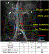
Regions of interest (ROIs) drawn for the quantitative assessment of both relative signal ratios and relative contrast ratios in major abdominal vascular segments are schematically shown on a coronal MIP image obtained using the VSS-prepared full angiogram
FIGURE 9
FIGURE 9
Averaged relative contrast ratios of artery-to-tissue at different vascular locations on VSS angiogram, SSI+VSS and VSI+VSS arteriograms (A), and artery-to-vein on SSI+VSS and VSI+VSS arteriograms, respectively (B)

Similar articles

Cited by

References

    1. Laissy JP, Trillaud H, Douek P. MR angiography: noninvasive vascular imaging of the abdomen. Abdom Imaging. 2002;27:488–506. - PubMed
    1. Hartung MP, Grist TM, François CJ. Magnetic resonance angiography: current status and future directions. J Cardiovasc Magn Reson. 2011;13:19. - PMC - PubMed
    1. Prince MR. Gadolinium-enhanced MR aortography. Radiology. 1994;191:155–164. - PubMed
    1. Prince MR, Narasimham DL, Stanley JC, et al. Breath-hold gadolinium-enhanced MR angiography of the abdominal aorta and its major branches. Radiology. 1995;197:785–792. - PubMed
    1. Hany TF, Debatin JF, Leung DA, Pfammatter T. Evaluation of the aortoiliac and renal arteries: comparison of breath-hold, contrast-enhanced, three-dimensional MR angiography with conventional catheter angiography. Radiology. 1997;204:357–362. - PubMed

Publication types