

Non-contrast-enhanced abdominal MRA at 3 T using velocity-selective pulse trains
- PMID: 32017173
- PMCID: PMC7263981
- DOI: 10.1002/mrm.28187
Non-contrast-enhanced abdominal MRA at 3 T using velocity-selective pulse trains
Abstract
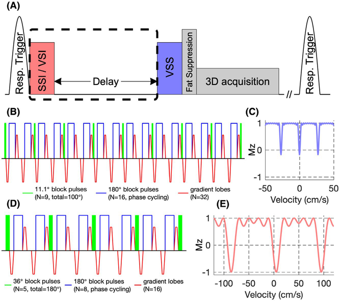
Purpose: Most existing non-contrast-enhanced methods for abdominal MR arteriography rely on a spatially selective inversion (SSI) pulse with a delay to null both static tissue and venous blood, and are limited to small spatial coverage due to the sensitivity to slow arterial inflow. Velocity-selective inversion (VSI) based approach has been shown to preserve the arterial blood inside the imaging volume at 1.5 T. Recently, velocity-selective saturation (VSS) pulse trains were applied to suppress the static tissue and have been combined with SSI pulses for cerebral MR arteriography at 3 T. The aim of this study is to construct an abdominal MRA protocol with large spatial coverage at 3 T using advanced velocity-selective pulse trains.
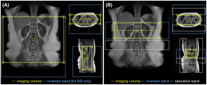
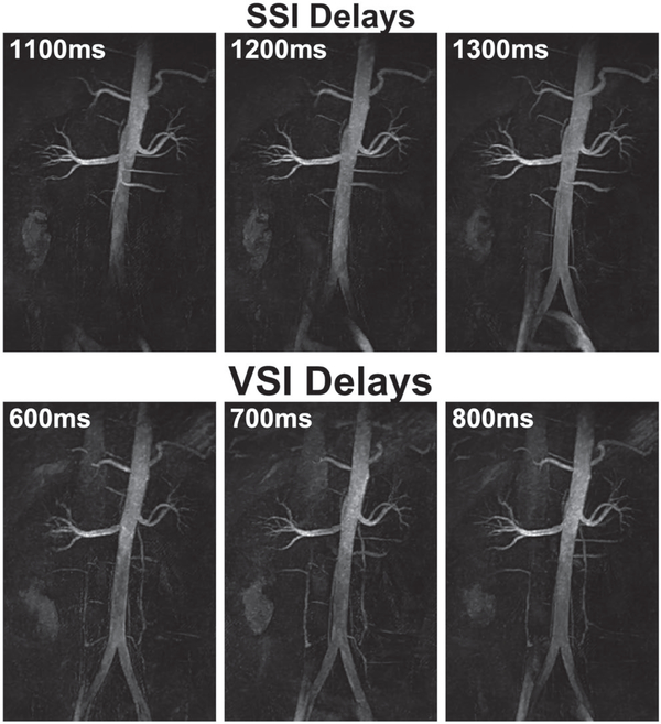
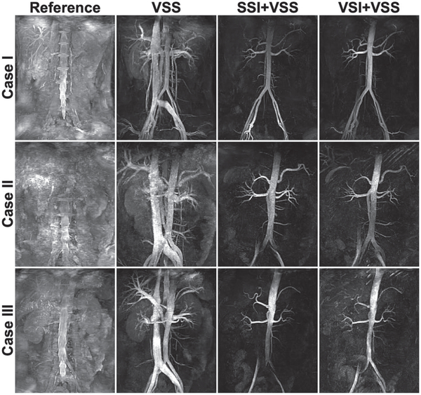
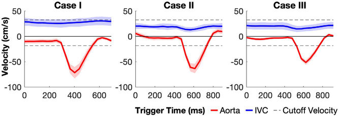
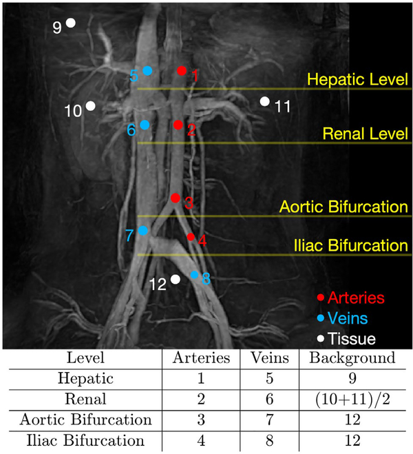
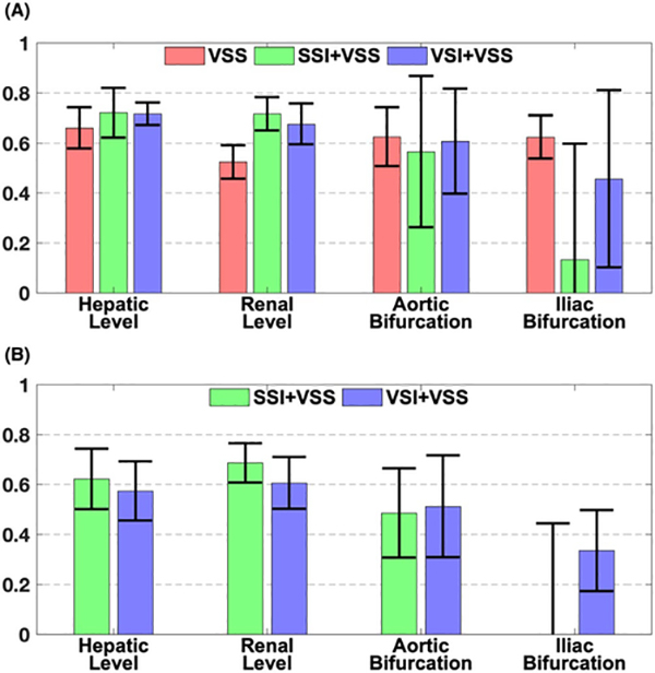
Methods: Multiple velocity-selective MRA protocols with different sequence modules and 3D acquisition methods were evaluated. Sequences using VSS only as well as SSI+VSS and VSI+VSS preparations were then compared among a group of healthy young and middle-aged volunteers. Using MRA without any preparations as reference, relative signal ratios and relative contrast ratios of different vascular segments were quantitatively analyzed.
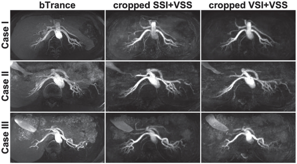
Results: Both SSI+VSS and VSI+VSS arteriograms achieved high artery-to-tissue and artery-to-vein relative contrast ratios above aortic bifurcation. The SSI+VSS sequence yielded lower signal at the bilateral iliac arteries than VSI+VSS, reflecting the benefit of the VSI preparation for imaging the distal branches.
Conclusion: The feasibility of noncontrast 3D MR abdominal arteriography was demonstrated on healthy volunteers using a combination of VSS pulse trains and SSI or VSI pulse.
Keywords: abdominal MRA; arteriography; non-contrast-enhanced MRA; velocity-selective pulse train.
© 2020 International Society for Magnetic Resonance in Medicine.
Figures

References
-
- Laissy JP, Trillaud H, Douek P. MR angiography: noninvasive vascular imaging of the abdomen. Abdom Imaging. 2002;27:488–506. - PubMed
-
- Prince MR. Gadolinium-enhanced MR aortography. Radiology. 1994;191:155–164. - PubMed
-
- Prince MR, Narasimham DL, Stanley JC, et al. Breath-hold gadolinium-enhanced MR angiography of the abdominal aorta and its major branches. Radiology. 1995;197:785–792. - PubMed
-
- Hany TF, Debatin JF, Leung DA, Pfammatter T. Evaluation of the aortoiliac and renal arteries: comparison of breath-hold, contrast-enhanced, three-dimensional MR angiography with conventional catheter angiography. Radiology. 1997;204:357–362. - PubMed
Publication types
MeSH terms
Substances
Grants and funding
LinkOut - more resources
Full Text Sources
