

Diagnosis efficacy of CEUS for hepatic inflammatory lesions
- PMID: 32017229
- PMCID: PMC7307339
- DOI: 10.1002/jcla.23231
Diagnosis efficacy of CEUS for hepatic inflammatory lesions
Abstract
Purpose: In this study, the efficacy of US/CEUS and clinicopathologic parameters in differential diagnosis of hepatic inflammatory lesions were evaluated.
Methods: This was a retrospective study in which CEUS imaging was performed on 182 patients. Among these patients, 44 patients had hepatic inflammatory lesions and 138 patients had malignant lesions. The ultrasound (US), CEUS, and clinicopathologic parameters with respect to differential diagnosis of hepatic inflammatory lesions were analyzed.
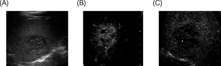
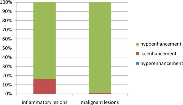
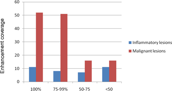
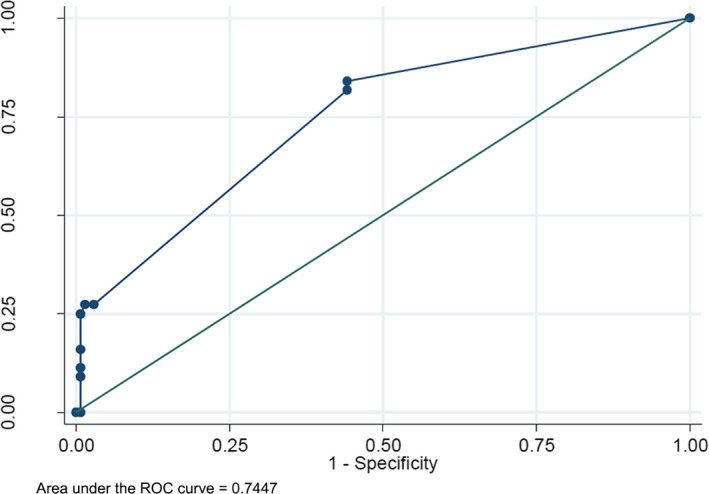
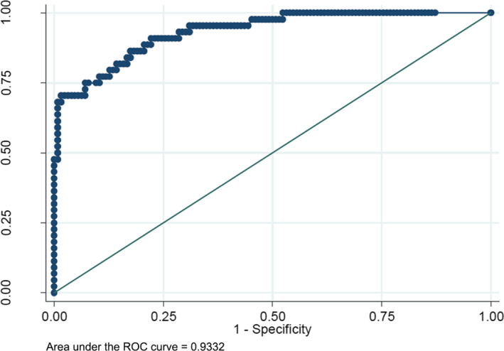
Results: Irregular lesion shape and unclear margin were commonly seen in hepatic inflammatory lesions by US/CEUS examination. Hypoenhancement in arterial phase (AP) and portal venous phase (PVP), and isoenhancement in delayed phase (DP) were more commonly found in inflammatory lesions rather than malignant lesions [9% (4/44), 68% (30/44), and 16% (7/44) vs 2% (3/138), 11% (15/138), 1% (1/138), respectively; P < .05]. The enhancement coverage was also a significant indicator for the differentiation of inflammatory lesions and malignant lesions (P < .05). History of hepatitis or cirrhosis, and higher serum alpha-fetoprotein (AFP) level were indicators for malignant lesions, while liver parasites and higher body temperature were indicators for inflammatory lesions. When the US/CEUS findings were combined with clinicopathologic parameters, the diagnostic accuracy of inflammatory lesions could reach 93.3%, with sensitivity, specificity, positive predictive value, and negative predictive value of 63.64%, 96.03%, 84.85%, and 88.32%, respectively.
Conclusion: The US/CEUS findings combined with clinical characteristics can accurately differentiate hepatic inflammatory lesions and malignant lesions. The results of study will improve the diagnostic confidence for hepatic inflammatory lesions.
Keywords: contrast-enhanced ultrasound imaging; differential diagnosis; hepatic inflammatory lesion.
© 2020 The Authors. Journal of Clinical Laboratory Analysis Published by Wiley Periodicals, Inc.
Figures
References
-
- Colmenero Jde D, Queipo‐Ortuño MI, Maria Reguera J, et al. Chronic hepatosplenic abscesses in Brucellosis. Clinico‐therapeutic features and molecular diagnostic approach. Diagn Microbiol Infect Dis. 2002;42:159‐167. - PubMed
-
- Dehner LP. The enigmatic inflammatory pseudotumours: the current state of our understanding, or misunderstanding. J Pathol. 2000;192:277‐279. - PubMed
-
- Joo YE, Kim HS, Choi SK, et al. Hemobilia caused by liver abscess due to intrahepatic duct stones. J Gastroenterol. 2003;38:507‐511. - PubMed
MeSH terms
Substances
Grants and funding
LinkOut - more resources
Full Text Sources
Medical
