. 2020 May;18(5):1027-1038.
doi: 10.1111/jth.14756. Epub 2020 Mar 5.

C-terminal residues of activated protein C light chain contribute to its anticoagulant and cytoprotective activities

Affiliations

C-terminal residues of activated protein C light chain contribute to its anticoagulant and cytoprotective activities

Atsuki Yamashita et al. J Thromb Haemost. 2020 May.

Abstract

Background: Activated protein C (APC) is an important homeostatic blood coagulation protease that conveys anticoagulant and cytoprotective activities. Proteolytic inactivation of factors Va and VIIIa facilitated by cofactor protein S is responsible for APC's anticoagulant effects, whereas cytoprotective effects of APC involve primarily the endothelial protein C receptor (EPCR), protease activated receptor (PAR)1 and PAR3.

Objective: To date, several binding exosites in the protease domain of APC have been identified that contribute to APC's interaction with its substrates but potential contributions of the C-terminus of the light chain have not been studied in detail.

Methods: Site-directed Ala-scanning mutagenesis of six positively charged residues within G142-L155 was used to characterize their contributions to APC's anticoagulant and cytoprotective activities.

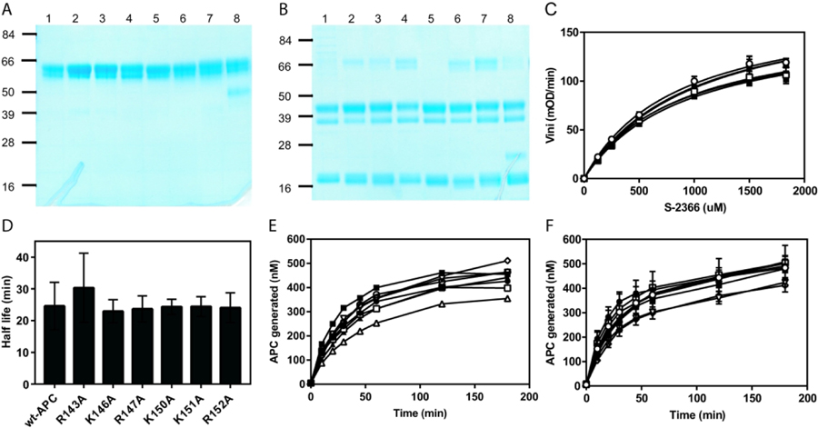
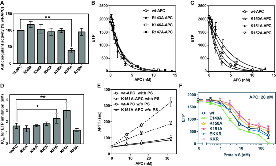
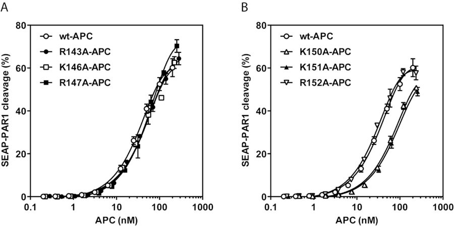
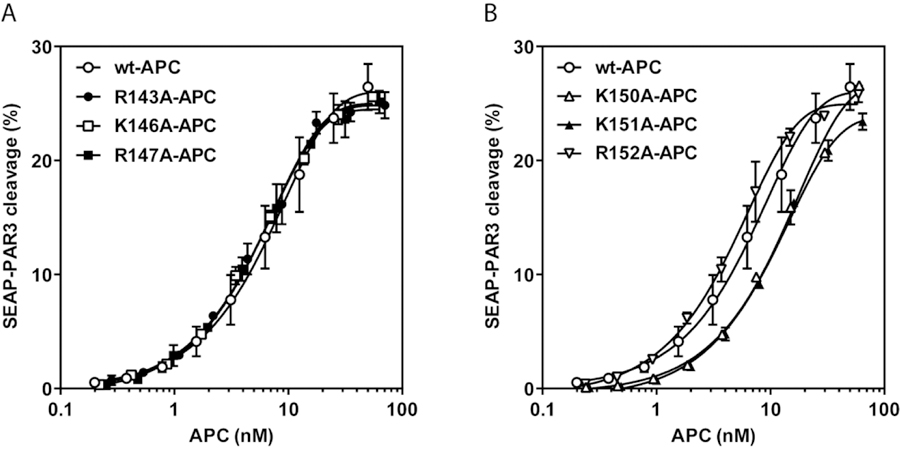
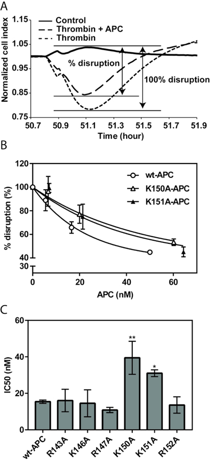
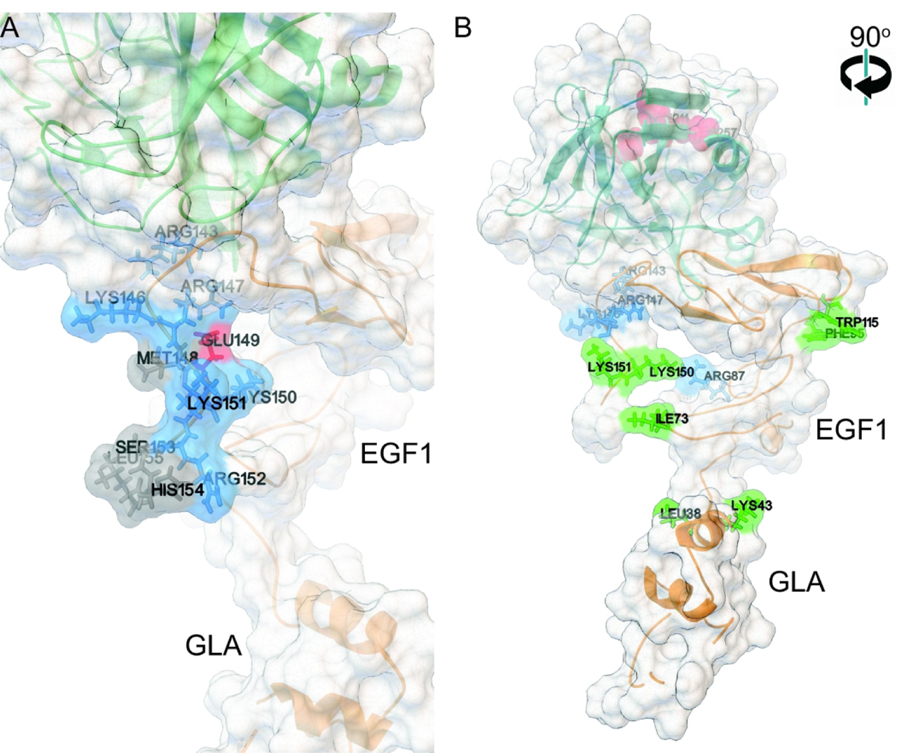
Results and conclusions: K151 was involved in protein S dependent-anticoagulant activity of APC with some contribution of K150. 3D structural analysis supported that these two residues were exposed in an extended protein S binding site on one face of APC. Both K150 and K151 were important for PAR1 and PAR3 cleavage by APC, suggesting that this region may also mediate interactions with PARs. Accordingly, APC's cytoprotective activity as determined by endothelial barrier protection was impaired by Ala substitutions of these residues. Thus, both K150 and K151 are involved in APC's anticoagulant and cytoprotective activities. The differential contribution of K150 relative to K151 for protein S-dependent anticoagulant activity and PAR cleavage highlights that binding exosites for protein S binding and for PAR cleavage in the C-terminal region of APC's light chain overlap.

Keywords: activated protein C; anticoagulant activity; cytoprotective activity; protease activated receptor; protein S.


Conflict of interest statement

CONFLICT OF INTEREST:

TSRI holds intellectual property rights related to some APC variants on which JHG and LOM are listed as inventors. The other authors declare that they have no conflicts of interest.

Figures

Figure 1.
Figure 1.. Characterization of protein C light chain mutants.
SDS-PAGE and Coomassie staining of protein C light chain mutants under non-reducing (A) and reducing (B) conditions. (wt (lane 1); R143A (lane 2); K146A (lane3); R147A (lane 4); E149A (lane5); K150A (lane 6); K151A (lane 7); R152A (lane 8)). (C) The cleavage rate of increasing concentrations of the small chromogenic substrate, S-2366, by wt-APC and APC mutants (10 nM). (D) Half-life of APC’s amidolytic activity in plasma. (E, F) Activation of protein C mutants by thrombin in the presence of 2 mM EDTA (E) or by thrombin and rabbit lung thrombomodulin in the presence of 3 mM CaCl2 (F). APC: wt (○), R143A (●), K146A (□), R147A (■), K150A (Δ), K151A (▲), R152A (▽), KKR150–152AAA (◊).
Figure 2.
Figure 2.. Anticoagulant activity of APC light chain mutants.
(A) Anticoagulant activity determined using activated partial thromboplastin time (APTT) assays. The anticoagulant activity was derived from APC dose-response curves using 0.5–32 nM APC with the activity of wt-APC was defined as 100%. (B, C) Inhibition of thrombin generation in normal plasma by wt-APC and APC mutants (B) R143A-APC, K146A-APC, R147A-APC and (C) K150A-APC, K151A-APC, R152A-APC. ETP denotes the endogenous thrombin potential. (D) APC concentrations of wt-APC and APC mutants required for half-maximal inhibition (IC50) of the ETP, calculated from the non-linear regression curves in panels B-C. (E) Effects of wt-APC and K151A-APC on APTT clotting times in protein S-depleted plasma with (dashed line) or without (solid line) reconstitution of protein S. (F) Inhibition of ETP by wt-APC, E149A-APC, K150A-APC, K151A-APC, EKKR149–152AAAA-APC and KKR150–152AAA-APC in protein S-depleted plasma reconstituted with an increasing concentration of protein S. Each data point represents the mean ± SD of 3 independent experiments. APC: wt (○), R143A (●), K146A (□), R147A (■), E149A (X), K150A (Δ), K151A (▲), R152A (▽), EKKR149–152AAAA (▼), KKR150–152AAA (◊).*P < 0.05; **P < 0.01.
Figure 3.
Figure 3.. PAR1 cleavage by APC light chain mutants.
APC-mediated cleavage of PAR1 was analyzed by the proteolytic release of SEAP from SEAP-PAR1-expressing HEK-293 cells in the presence of co-transfected wild-type (wt)-EPCR. (A) Dose response of PAR1 cleavage by wt-APC (○), R143A-APC (●), K146A-APC (□) and R147A-APC (■). (B) Dose response of PAR1 cleavage by wt-APC (○), K150A-APC (Δ), K151A-APC (▲), R152A-APC (▽). PAR1 cleavage was expressed as a percentage of the total available SEAP-PAR1 on the cells. Experiments were performed in duplicate and data points represent the mean ± SD.
Figure 4.
Figure 4.. PAR3 cleavage by APC light chain mutants.
APC-mediated cleavage of PAR3 was analyzed by the proteolytic release of SEAP from SEAP-PAR3-expressing HEK-293 cells in the presence of co-transfected wild-type (wt)-EPCR. (A) Dose response of PAR3 cleavage by wt-APC (○), R143A-APC (●), K146A-APC (□) and R147A-APC (■). (B) Dose response of PAR3 cleavage by wt-APC (○), K150A-APC (Δ), K151A-APC (▲), R152A-APC (▽). PAR3 cleavage was expressed as a percentage of the total available SEAP-PAR3 on the cells. Experiments were performed in duplicate and data points represent the mean ± SD.
Figure 5.
Figure 5.. Endothelial barrier protection by APC light chain mutants.
Protection of endothelial barrier function by wt-APC and APC mutants against thrombin-induced permeability (2 nM). (A) Barrier function of EA.hy926 endothelial cells was assessed by ECIS in real-time by monitoring changes in TER. Results are expressed as percentage of maximal barrier disruption induced by thrombin in the absence of APC set as 100%. (B) Concentration-dependent endothelial barrier protection by wt-APC (○), K150A-APC (Δ), K151A-APC (▲). (C) The concentration of wt-APC and APC mutants required for half-maximal (IC50) endothelial barrier protection. Data points represent the mean ± SD of 3 independent experiments. *P < 0.05, **P < 0.01 versus wt-APC.
Figure 6.
Figure 6.. Structure of the C-terminus of the APC light chain obtained by molecular dynamics.
(A) Closeup of the C-terminus of the APC light chain in the conformation observed most often during the MD simulations. (B) An overview (rotated by 90 degrees along the vertical axis compared to panel A) of the same APC conformation with the light chain ribbon diagram in gold color and the protease domain ribbon in dark green color. The atoms of the catalytic triad are displayed as red van der Waals spheres to help with the overall orientation of the complex. A semi-transparent molecular surface is displayed to convey the overall shape of the assembly [63]. Mutations of the amino acids L38, K43, I73, K150, K151, F95 and W115, shown in green, reduce the enhancement of APC’s anticoagulant activity by protein S. They delinate a groove where protein S can interact with APC. The mutations of amino acids R87, K146 R143 and R147, shown in blue, do not affect anticoagulant acitivty enhancement by protein S and are located away from the hypothetical protein S-binding groove. The nature of the groove and the relative position of the blue and green residues (B) can best be seen in the short video (Supporting Movie S1) provided in the suporting information section.

Similar articles

Cited by

References

    1. O’Donnell JS, O’Sullivan JM, Preston RJS. Advances in understanding the molecular mechanisms that maintain normal haemostasis. Br J Haematol. 2019; 186: 24–36. - PubMed
    1. Dahlback B, Villoutreix BO. Regulation of blood coagulation by the protein C anticoagulant pathway: novel insights into structure-function relationships and molecular recognition. Arterioscler Thromb Vasc Biol. 2005; 25: 1311–20. - PubMed
    1. Wildhagen KC, Lutgens E, Loubele ST, ten Cate H, Nicolaes GA. The structure-function relationship of activated protein C. Lessons from natural and engineered mutations. Thromb Haemost. 2011; 106: 1034–45. - PubMed
    1. Mosnier LO, Griffin JH. Protein C, protein S, thrombomodulin and the endothelial protein C receptor pathways In: Marder VJ, Aird WC, Bennett JS, Schulman S, White GC, Colman RW, eds. Hemostasis and Thrombosis: Basic Principles and Clinical Practice, 6th edn. Philadelphia: Lippincott Williams & Wilkins, 2013, 300–13.
    1. Beckmann RJ SR, Santerre RF, Plutzky J, Crabtree GR, Long GL. The structure and evolution of a 461 amino acid human protein C precursor and its messenger RNA, based upon the DNA sequence of cloned human liver cDNAs. Nucleic Acids Res. 1985; 13: 5233–47. - PMC - PubMed

Publication types