

Alteration of cell junctions during viral infection
- PMID: 32017415
- PMCID: PMC7049484
- DOI: 10.1111/1759-7714.13344
Alteration of cell junctions during viral infection
Abstract
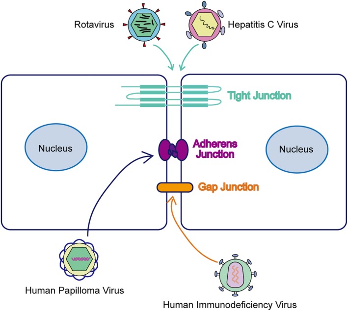
Cell junctions serve as a protective barrier for cells and provide an important channel for information transmission between cells and the surrounding environment. Viruses are parasites that invade and commandeer components of host cells in order to survive and replicate, and they have evolved various mechanisms to alter cell junctions to facilitate viral infection. In this review, we examined the current state of knowledge on the action of viruses on host cell junctions. The existing evidence suggests that targeting the molecules involved in the virus-cell junction interaction can prevent the spread of viral diseases.
Keywords: Adherens junction; gap junction; infection; tight junction; virus.
© 2020 The Authors. Thoracic Cancer published by China Lung Oncology Group and John Wiley & Sons Australia, Ltd.
Figures

 The purple cell) HIV infected cell, (The green cells
 ) Uninfected cell, (
 The yellow gap junctions) Normal gap junction, and (
 The purple gap junctions) HIV “Hijacked” gap junction.
References
Publication types
MeSH terms
Grants and funding
LinkOut - more resources
Full Text Sources
Medical
Miscellaneous
