

Thermoresponsive Inverted Colloidal Crystal Hydrogel Scaffolds for Lymphoid Tissue Engineering
- PMID: 32017462
- PMCID: PMC7103457
- DOI: 10.1002/adhm.201901556
Thermoresponsive Inverted Colloidal Crystal Hydrogel Scaffolds for Lymphoid Tissue Engineering
Abstract
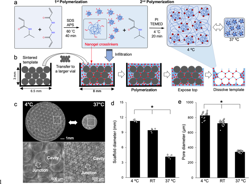
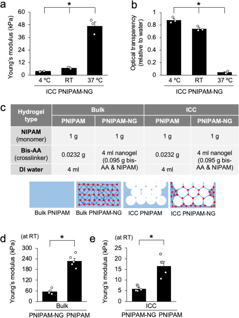
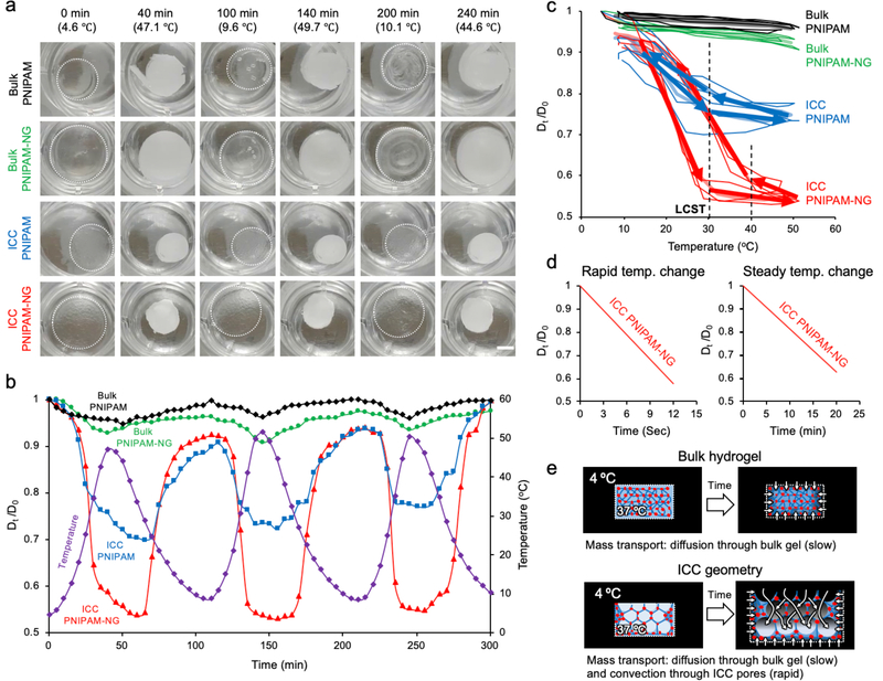
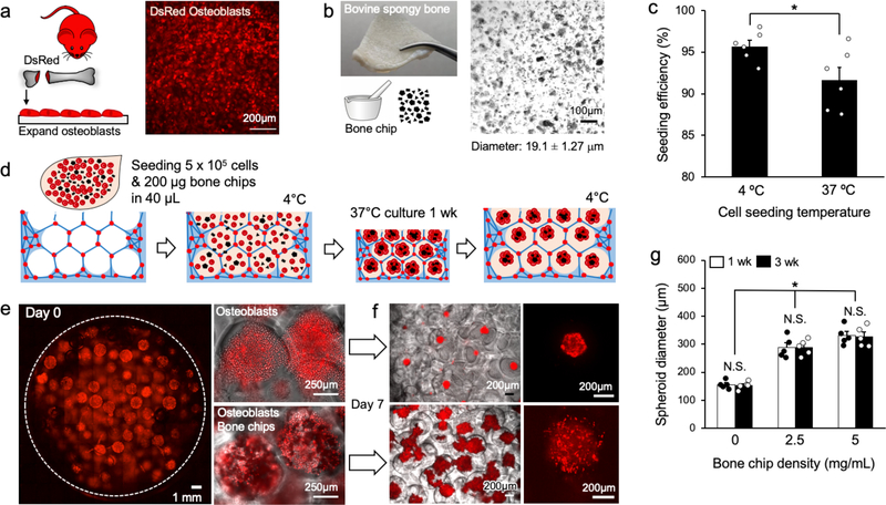
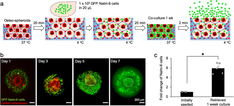
Inverted colloidal crystal (ICC) hydrogel scaffolds represent unique opportunities in modeling lymphoid tissues and expanding hematopoietic-lymphoid cells. Fully interconnected spherical pore arrays direct the formation of stromal networks and facilitate interactions between stroma and hematopoietic-lymphoid cells. However, due to the intricate architecture of these materials, release of expanded cells is restricted and requires mechanical disruption or chemical dissolution of the hydrogel scaffold. One potent biomaterials strategy to release pore-entrapped hematopoietic-lymphoid cells without breaking the scaffolds apart is to transiently increase the dimensions of these materials using stimuli-responsive polymers. Having this mindset, thermoresponsive ICC scaffolds that undergo rapid (<1 min) and substantial (>300%) diameter change over a physiological temperature range (4-37 °C) by using poly(N-isopropylacrylamide) (PNIPAM) with nanogel crosslinkers is developed. For a proof-of-concept study, the stromal niche by creating osteospheroids, aggregates of osteoblasts, and bone chips is first replicated, and subsequently Nalm-6 model hematopoietic-lymphoid cells are introduced. A sixfold increase in cell count is harvested when ICC hydrogel scaffolds are expanded without termination of the established 3D stromal cell culture. It is envisioned that thermoresponsive ICC hydrogel scaffolds will enable for scalable and sustainable ex vivo expansion of hematopoietic-lymphoid cells.
Keywords: PNIPAM; hydrogel scaffolds; inverted colloidal crystals; lymphoid tissue; stimuli-responsive materials.
© 2020 WILEY-VCH Verlag GmbH & Co. KGaA, Weinheim.
Conflict of interest statement
Conflict of Interest
The authors declare no conflict of interest.
Figures
Similar articles
-
Fabrication of Bioactive Inverted Colloidal Crystal Scaffolds Using Expanded Polystyrene Beads.Tissue Eng Part C Methods. 2020 Mar;26(3):143-155. doi: 10.1089/ten.TEC.2019.0333. Epub 2020 Mar 6. Tissue Eng Part C Methods. 2020. PMID: 32031058 Free PMC article.
-
Electrospun thermosensitive hydrogel scaffold for enhanced chondrogenesis of human mesenchymal stem cells.Acta Biomater. 2018 Jan 15;66:166-176. doi: 10.1016/j.actbio.2017.11.020. Epub 2017 Nov 8. Acta Biomater. 2018. PMID: 29128540
-
Mechanisms of pore formation in hydrogel scaffolds textured by freeze-drying.Acta Biomater. 2019 Aug;94:195-203. doi: 10.1016/j.actbio.2019.05.070. Epub 2019 May 30. Acta Biomater. 2019. PMID: 31154055
-
Biocompatibility of hydrogel-based scaffolds for tissue engineering applications.Biotechnol Adv. 2017 Sep;35(5):530-544. doi: 10.1016/j.biotechadv.2017.05.006. Epub 2017 May 27. Biotechnol Adv. 2017. PMID: 28558979 Review.
-
Macroporous Hydrogel Scaffolds for Three-Dimensional Cell Culture and Tissue Engineering.Tissue Eng Part B Rev. 2017 Oct;23(5):451-461. doi: 10.1089/ten.TEB.2016.0465. Epub 2017 Feb 3. Tissue Eng Part B Rev. 2017. PMID: 28067115 Review.
Cited by
-
Urine-derived stem cells: Promising advancements and applications in regenerative medicine and beyond.Heliyon. 2024 Mar 10;10(6):e27306. doi: 10.1016/j.heliyon.2024.e27306. eCollection 2024 Mar 30. Heliyon. 2024. PMID: 38509987 Free PMC article. Review.
-
Lymphatic Tissue and Organ Engineering for In Vitro Modeling and In Vivo Regeneration.Cold Spring Harb Perspect Med. 2022 Mar 14;12(9):a041169. doi: 10.1101/cshperspect.a041169. Online ahead of print. Cold Spring Harb Perspect Med. 2022. PMID: 35288402 Free PMC article.
-
Bone Marrow Adipocytes Contribute to Tumor Microenvironment-Driven Chemoresistance via Sequestration of Doxorubicin.Cancers (Basel). 2023 May 12;15(10):2737. doi: 10.3390/cancers15102737. Cancers (Basel). 2023. PMID: 37345073 Free PMC article.
-
Recent advances in engineering hydrogels for niche biomimicking and hematopoietic stem cell culturing.Front Bioeng Biotechnol. 2022 Nov 24;10:1049965. doi: 10.3389/fbioe.2022.1049965. eCollection 2022. Front Bioeng Biotechnol. 2022. PMID: 36507253 Free PMC article. Review.
-
Poly(N-isopropylacrylamide)-Based Hydrogels for Biomedical Applications: A Review of the State-of-the-Art.Gels. 2022 Jul 20;8(7):454. doi: 10.3390/gels8070454. Gels. 2022. PMID: 35877539 Free PMC article. Review.
References
-
- Lee J; Cuddihy MJ; Kotov NA, Tissue Eng Pt B-Rev 2008, 14 (1), 61–86. - PubMed
-
- Hussey GS; Dziki JL; Badylak SF, Nat Rev Mater 2018, 3 (7), 159–173.
-
- Lee J; Kotov NA, Small 2009, 5 (9), 1008–1013. - PubMed
-
- Stachowiak AN; Irvine DJ, Journal of Biomedical Materials Research Part A 2008, 85a (3), 815–828. - PubMed
Publication types
MeSH terms
Substances
Grants and funding
LinkOut - more resources
Full Text Sources
