. 2020 Feb 21;15(2):325-333.
doi: 10.1021/acschembio.9b00963. Epub 2020 Feb 10.

Structure-Guided Design and In-Cell Target Profiling of a Cell-Active Target Engagement Probe for PARP Inhibitors

Affiliations

Structure-Guided Design and In-Cell Target Profiling of a Cell-Active Target Engagement Probe for PARP Inhibitors

Ryan T Howard et al. ACS Chem Biol. .

Abstract

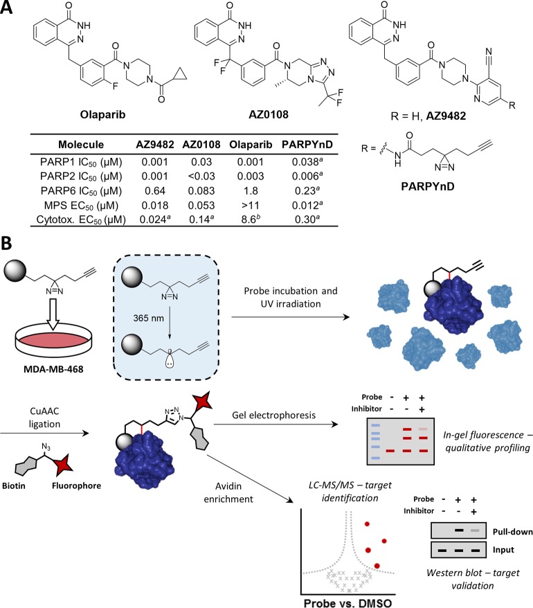
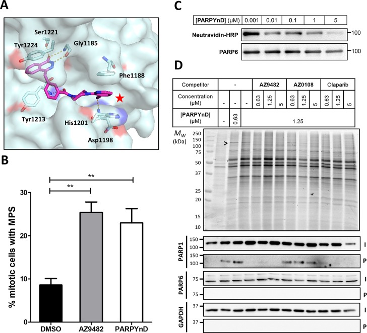
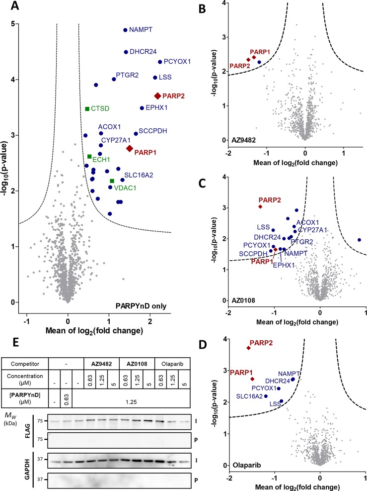
Inhibition of the poly(ADP-ribose) polymerase (PARP) family of enzymes has become an attractive therapeutic strategy in oncology and beyond; however, chemical tools to profile PARP engagement in live cells are lacking. Herein, we report the design and application of PARPYnD, the first photoaffinity probe (AfBP) for PARP enzymes based on triple PARP1/2/6 inhibitor AZ9482, which induces multipolar spindle (MPS) formation in breast cancer cells. PARPYnD is a robust tool for profiling PARP1/2 and is used to profile clinical PARP inhibitor olaparib, identifying several novel off-target proteins. Surprisingly, while PARPYnD can enrich recombinant PARP6 spiked into cellular lysates and inhibits PARP6 in cell-free assays, it does not label PARP6 in intact cells. These data highlight an intriguing biomolecular disparity between recombinant and endogenous PARP6. PARPYnD provides a new approach to expand our knowledge of the targets of this class of compounds and the mechanisms of action of PARP inhibitors in cancer.


Conflict of interest statement

The authors declare the following competing financial interest(s): This work was partially funded by AstraZeneca, and P.H., P.P., J.S.S., and J.W.J. are all employees of AstraZeneca. E.W.T. is a Director and shareholder of Myricx Pharma Ltd.

Figures

Figure 1
Figure 1
A photoaffinity-based probe (AfBP) PARPYnD was designed and synthesized for target profiling of AZ9482 and AZ0108. (A) Top: structures of clinical PARP1/2 inhibitor olaparib and MPS-inducing agents AZ0108 and AZ9482, the latter of which was diversified into the AfBP used in this study. Bottom: table showing the biochemical and biophysical parameters associated with olaparib, AZ9482, AZ0108, and PARPYnD., Table footnote a: data generated in this work, experiments performed in triplicate (±SEM); graphical analysis can be seen in Figure 2, and Supporting Information Figures S1 and S2 for multipolar spindle (MPS) induction data, PARP binding data, and cytotoxicity data (MDA-MB-468 cells), respectively. Table footnote b: cytotoxicity value represents a GI50 value previously generated in MDA-MB-468 cells. (B) Schematic of the photoaffinity labeling (PAL) workflow used for target profiling. The gray ball represents the recognition element of the probe that is designed based on the parent compound.
Figure 2
Figure 2
Validation of PARPYnD as a suitable probe for parent molecule profiling. (A) Crystal structure of parent molecule AZ9482 bound in the NAD+-binding pocket of PARP5a (PDB ID: 5ECE), with key interactions highlighted in orange. The red star highlights the solvent exposed position at which modification is expected to minimally perturb the binding of a probe into this pocket of the PARP enzymes. (B) Quantification of the percentage of mitotic cells with MPS phenotype (>2 spindle poles per cell) after treatment with AZ9482 (N = 2) and PARPYnD (N = 2), both at 41 nM, versus DMSO (N = 4); double asterisk (**) represents raw P value <0.001 in unpaired Student’s t test; raw data found in the Supporting Information, Extended Data S1. (C) PARP6 activity assay: recombinant GST-tagged PARP6 was incubated with biotinylated NAD+ and varying concentrations of PARPYnD. GST-PARP6 auto-MARylation was measured by immunoblotting against NeutrAvidin-HRP; decreased signal with increasing PARPYnD concentration indicated catalytic inhibition of PARP6. (D) Gel and Western blot analysis of live cells labeled with PARPYnD and ligated to AzTB (Figure S3A) with/without cotreatment with various competitor molecules. In-gel fluorescence was used to qualitatively assess TAMRA-tagged proteins, and streptavidin-based enrichment and immunoblot analysis was used to validate specific targets. I = input (whole lysate); P = pull down (enriched fraction); > indicates competition seen on gel, validated as PARP1 by immunoblot. Uncropped gels and immunoblots associated with all figures can be found in Figure S7.
Figure 3
Figure 3
Target engagement profiles of PARPYnD, AZ9482, AZ0108, and olaparib. (A–D) Proteomics analysis of live cells labeled with PARPYnD and ligated to AzRB (Figure S3B) with/without cotreatment with parent competitor molecules. Tagged proteins were enriched on NeutrAvidin agarose, digested into peptides, and tandem mass tag (TMT) labeled for identification and quantification by LC-MS/MS. Volcano plots demonstrate enrichment (x axis) of one sample versus another and the associated significance (y axis), determined by pairwise Student’s t test (cut off: A, S0 = 0.1, false discovery rate (FDR) = 5%; B–D, S0 = 0.1, FDR = 15%). Red diamonds = PARP family, blue dots = other significantly enriched/depleted hits, green squares = known background photocrosslinking binders, gray dots = nonsignificant proteins. Significant hits are annotated with their gene names only when they are appear significantly enriched/depleted across more than one pairwise comparison, with the exception of the known background binders. Larger, fully annotated plots can be found in Figure S5A–D, and complete raw data can be found in the Supporting Information, Extended Data S2. (A) PARPYnD (1 μM) versus DMSO. (B) PARPYnD (1 μM) and AZ9482 (5 μM) versus PARPYnD (1 μM) only. (C) PARPYnD (1 μM) and AZ0108 (5 μM) versus PARPYnD (1 μM) only. (D) PARPYnD (1 μM) and olaparib (5 μM) versus PARPYnD (1 μM) only. (E) Transient overexpression of FLAG-PARP6 and attempted enrichment of the overexpressed protein and identification by Western blot. I = input (whole lysate); P = pull down (enriched fraction).
Figure 4
Figure 4
Successful engagement of recombinant but not endogenous PARP6 in lysate-based crosslinking experiments. In-gel fluorescence (top) and immunoblot (bottom) analysis of lysates labeled with PARPYnD and ligated to AzTB with/without cotreatment with AZ0108. Left panel—lysates were spiked with recombinant GST-PARP6 before treatment. Ligated samples were further enriched on streptavidin beads and all samples analyzed as above: > GST-PARP6 (98 kDa); * PARP6 (71 kDa). More recombinant protein is labeled/enriched compared to vehicle (DMSO) control, where residual signal can be observed through nonspecific interaction of the recombinant protein with the beads. No enrichment of endogenous PARP6 is observed.

References

    1. Gibson B. A.; Kraus W. L. (2012) New insights into the molecular and cellular functions of poly(ADP-ribose) and PARPs. Nat. Rev. Mol. Cell Biol. 13, 411–424. 10.1038/nrm3376. - DOI - PubMed
    1. Vyas S.; Matic I.; Uchima L.; Rood J.; Zaja R.; Hay R. T.; Ahel I.; Chang P. (2014) Family-wide analysis of poly(ADP-ribose) polymerase activity. Nat. Commun. 5, 4426.10.1038/ncomms5426. - DOI - PMC - PubMed
    1. Yang C.-S.; Jividen K.; Spencer A.; Dworak N.; Ni L.; Oostdyk L. T.; Chatterjee M.; Kuśmider B.; Reon B.; Parlak M.; Gorbunova V.; Abbas T.; Jeffery E.; Sherman N. E.; Paschal B. M. (2017) Ubiquitin Modification by the E3 Ligase/ADP-Ribosyltransferase Dtx3L/Parp9. Mol. Cell 66, 503–516. 10.1016/j.molcel.2017.04.028. - DOI - PMC - PubMed
    1. Liu C.; Vyas A.; Kassab M. A.; Singh A. K.; Yu X. (2017) The role of poly ADP-ribosylation in the first wave of DNA damage response. Nucleic Acids Res. 45, 8129–8141. 10.1093/nar/gkx565. - DOI - PMC - PubMed
    1. Kraus W. L.; Hottiger M. O. (2013) PARP-1 and gene regulation: Progress and puzzles. Mol. Aspects Med. 34, 1109–1123. 10.1016/j.mam.2013.01.005. - DOI - PubMed

Publication types

MeSH terms