. 2020 Oct 1;49(7):20190445.
doi: 10.1259/dmfr.20190445. Epub 2020 Feb 6.

Ultrasonography for diagnosis of peri-implant diseases and conditions: a detailed scanning protocol and case demonstration

Affiliations

Ultrasonography for diagnosis of peri-implant diseases and conditions: a detailed scanning protocol and case demonstration

Hsun-Liang Chan et al. Dentomaxillofac Radiol. .

Abstract

Objectives: Ultrasonography has shown its promising diagnostic value in dental implant imaging research in the three treatment phases, namely, planning, intraoperative, and postoperative phase. With increasing awareness of peri-implant diseases and a lack of an efficient diagnostic method, the aim is to propose ultrasound imaging as a potential solution by providing a detailed scanning protocol and case demonstration.

Methods: Ultrasound device specification and the setup for optimizing peri-implant tissue imaging was described. Two useful imaging modes, viz. B-mode and color flow, were introduced. Important anatomical structures for accurate diagnosis of peri-implant diseases were illustrated. Finally, a detailed scanning sequence was proposed.

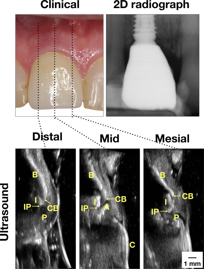
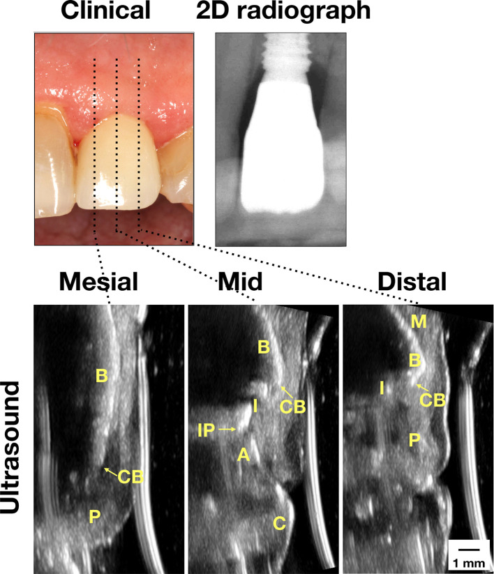
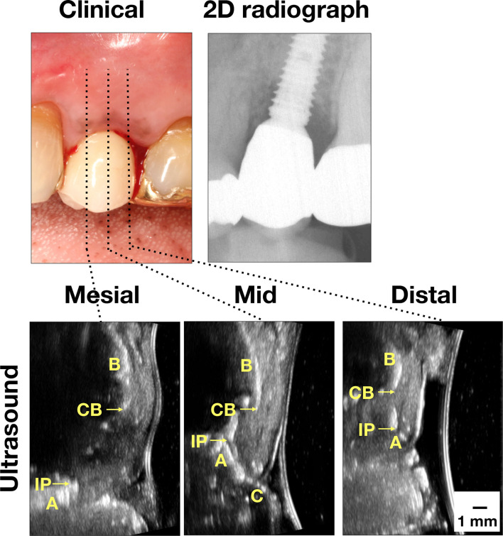
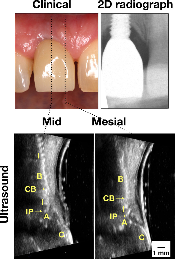
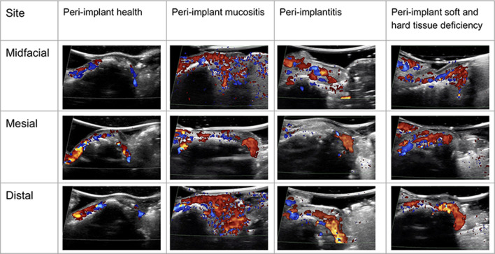
Results: Ultrasound images were acquired on live humans to exemplify the four peri-implant diseases and conditions, endorsed by the 2017 World Workshop organized by the American Academy of Periodontology and the European Federation of Periodontology. Ultrasound can provide not only cross-sectional anatomical images but also functional images (color flow images) that may be useful for evaluating the degree of peri-implant tissue inflammation.

Conclusions: High-frequency ultrasonography could be another cross-sectional imaging modality in adjunct to radiographs for diagnosing imminent peri-implant diseases and conditions that negatively influence quality of life of millions of patients with implants. This case study provides a framework for future related research work and clinical scanning guidelines.

Keywords: alveolar bone; cone-beam computed tomography; dental Implants; peri-implantitis; soft tissue; ultrasonography.


Figures

Figure 1.
Figure 1.
Case demonstration of a clinically healthy implant. Clinically, there are no signs of inflammation. Minimal bone loss was found on the peri-apical film. Ultrasound images on the mesial, mid-facial, and distal sites were presented. In the mid-facial image, the crown (C) surface is a bright line, connected to the abutment (A) and then the implant (I). The implant is readily identifiable because of the presence of the comet-tail artifact behind the implant. The bone surface (B) is yet another bright curved line. The junction between B and I is the CB. The junction between I and A is the IP. In the mesial and distal images, the bone and implant delineations are interpreted in the same way as in the mid-facial image. Additionally, the interdental papilla (P) is the structure between CB and the soft tissue surface. There is minimal implant fixture exposure, indicating normal bone level. CB,crestal bone; IP, implant platform.
Figure 2.
Figure 2.
Case demonstration of an implant with peri-implant mucositis. Clinically, the mucosa swelled and bled upon probing. Normal bone level was found on the peri-apical film. Ultrasound images on the mesial, mid-facial, and distal sites showed normal bone level as well. 2D, two-dimensional; A, abutment; B, bone; C, crown; CB, crestal bone; I, implant; IP, implant platform; M, mucosa; p, papilla).
Figure 3.
Figure 3.
Case demonstration of an implant with peri-implantitis. Clinically, there were signs of inflammation, including bleeding on probing. Bone loss was obvious on the mesial site on the peri-apical film. Ultrasound images also showed bone loss. 2D, two-dimensional; A, abutment; B, bone; C, crown; CB, crestal bone; I, implant; IP, implant platform; M, mucosa; p, papilla).
Figure 4.
Figure 4.
Case demonstration of an implant with hard and soft tissue deficiency. Clinically, there was slight inflammation. Some bone loss was found on the peri-apical film. Ultrasound images clearly showed thin mucosa and crestal bone, indicative of tissue deficiency. Implant exposure on the mid-facial view was evident. 2D, two-dimensional; A, abutment; B, bone; C, crown; CB, crestal bone; I, implant; IP, implant platform; M, mucosa; p, papilla.
Figure 5.
Figure 5.
Color flow images of the four representative cases. The color intensity and extent within soft tissues is less in the peri-implant healthy case, compared to the other disease cases. Color follow may be used to evaluate the degree of inflammation.

References

    1. Bhaskar V, Chan H-L, MacEachern M, Kripfgans OD. Updates on ultrasound research in implant dentistry: a systematic review of potential clinical indications. Dentomaxillofac Radiol 2018; 47: 20180076. doi: 10.1259/dmfr.20180076 - DOI - PMC - PubMed
    1. Chan H-L, Misch K, Wang H-L. Dental imaging in implant treatment planning. Implant Dent 2010; 19: 288–98. doi: 10.1097/ID.0b013e3181e59ebd - DOI - PubMed
    1. Schwarz F, Derks J, Monje A, Wang H-L, Peri-implantitis WHL. Peri-Implantitis. J Periodontol 2018; 89(Suppl 1): S267–90. doi: 10.1002/JPER.16-0350 - DOI - PubMed
    1. Derks J, Tomasi C. Peri-Implant health and disease. a systematic review of current epidemiology. J Clin Periodontol 2015; 42(Suppl 16): S158–71. doi: 10.1111/jcpe.12334 - DOI - PubMed
    1. Chan H-L, Lin G-H, Suarez F, MacEachern M, Wang H-L. Surgical management of peri-implantitis: a systematic review and meta-analysis of treatment outcomes. J Periodontol 2014; 85: 1027–41. doi: 10.1902/jop.2013.130563 - DOI - PubMed

Substances