

CT Imaging Features of 2019 Novel Coronavirus (2019-nCoV)
- PMID: 32017661
- PMCID: PMC7194022
- DOI: 10.1148/radiol.2020200230
CT Imaging Features of 2019 Novel Coronavirus (2019-nCoV)
Abstract
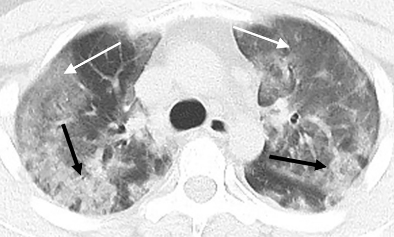
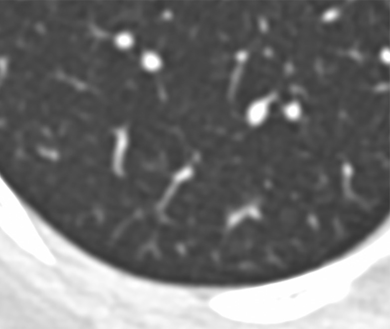
In this retrospective case series, chest CT scans of 21 symptomatic patients from China infected with the 2019 novel coronavirus (2019-nCoV) were reviewed, with emphasis on identifying and characterizing the most common findings. Typical CT findings included bilateral pulmonary parenchymal ground-glass and consolidative pulmonary opacities, sometimes with a rounded morphology and a peripheral lung distribution. Notably, lung cavitation, discrete pulmonary nodules, pleural effusions, and lymphadenopathy were absent. Follow-up imaging in a subset of patients during the study time window often demonstrated mild or moderate progression of disease, as manifested by increasing extent and density of lung opacities.
© RSNA, 2020.
Figures


References
-
- Novel Coronavirus – China. World Health Organization. https://www.who.int/csr/don/12-january-2020-novel-coronavirus-china/en/. Published January 12, 2020.
-
- Novel Coronavirus (2019-nCoV). World Health Organization. https://www.who.int/emergencies/diseases/novel-Coronavirus-2019. Published January 7, 2020.
-
- Summary of probable SARS cases with onset of illness from 1 November 2002 to 31 July 2003. World Health Organization. https://www.who.int/csr/sars/country/table2004_04_21/en/. Published April 21, 2004.
-
- Middle East respiratory syndrome Coronavirus (MERS-CoV). World Health Organization. https://www.who.int/emergencies/mers-cov/en/.
-
- Situation report - 8. World Health Organization. https://www.who.int/docs/default-source/Coronaviruse/situation-reports/2.... Published January 28, 2020.
MeSH terms
LinkOut - more resources
Full Text Sources
Other Literature Sources
Medical
