Review
. 2021 Jul:475:165-180.
doi: 10.1016/j.ydbio.2020.01.012. Epub 2020 Feb 1.

The role of cell lineage in the development of neuronal circuitry and function

Affiliations
Review

The role of cell lineage in the development of neuronal circuitry and function

Volker Hartenstein et al. Dev Biol. 2021 Jul.

Abstract

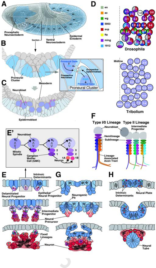
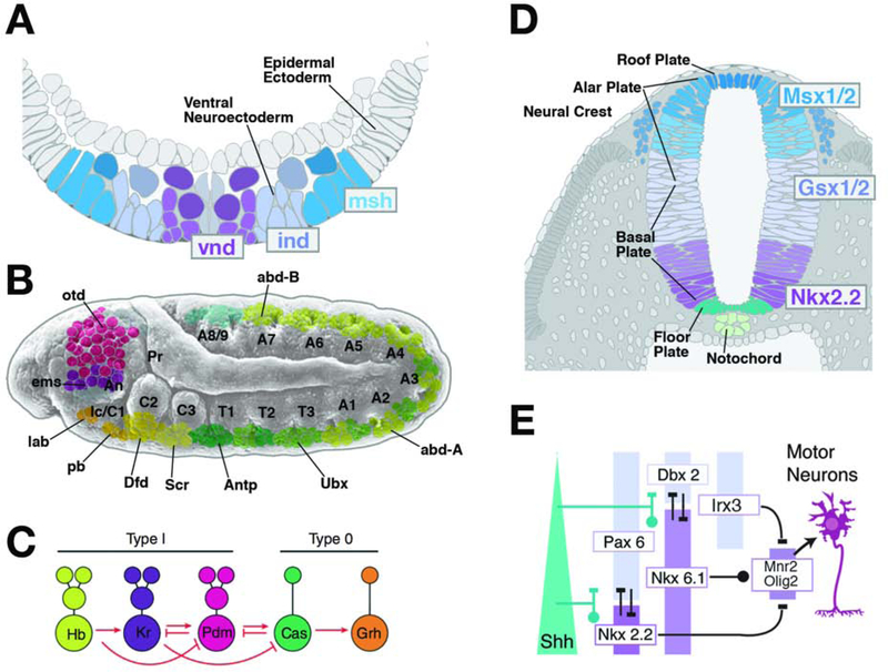
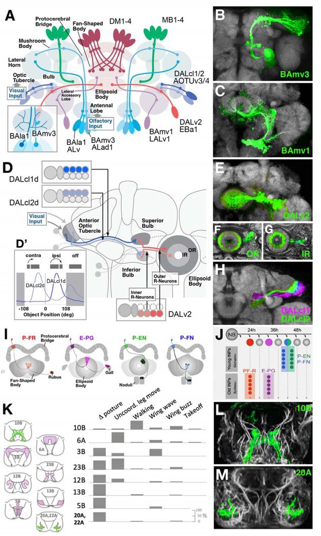
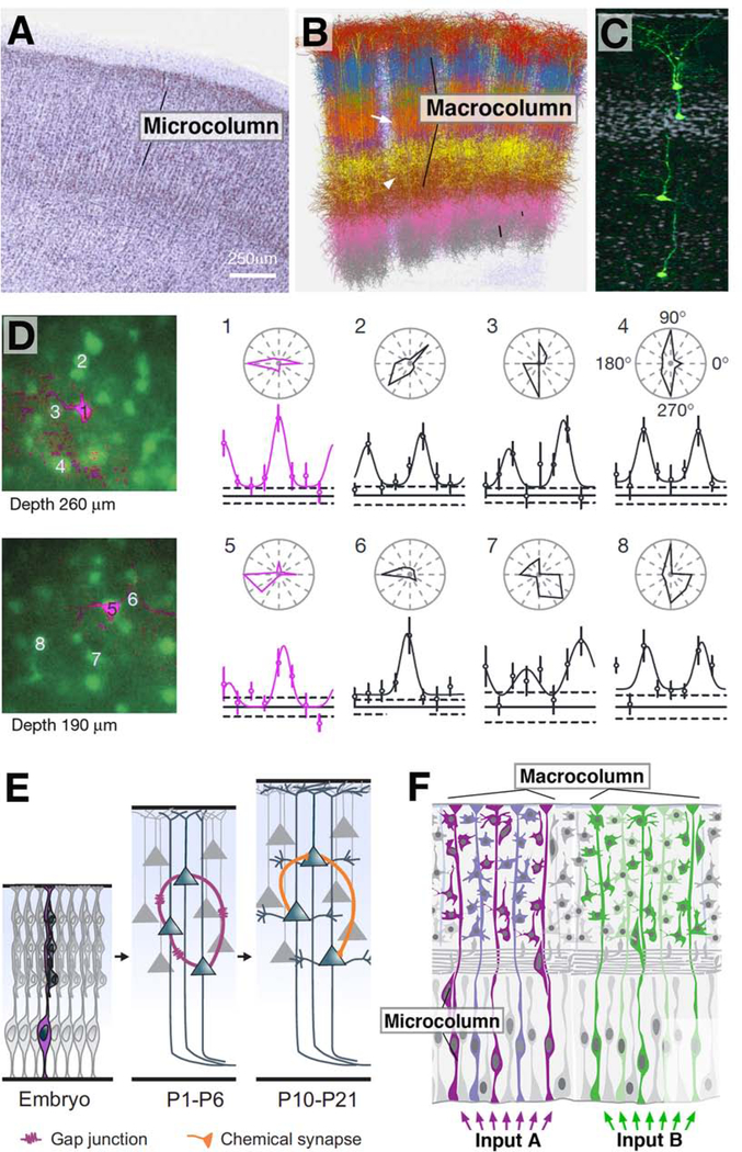
Complex nervous systems have a modular architecture, whereby reiterative groups of neurons ("modules") that share certain structural and functional properties are integrated into large neural circuits. Neurons develop from proliferating progenitor cells that, based on their location and time of appearance, are defined by certain genetic programs. Given that genes expressed by a given progenitor play a fundamental role in determining the properties of its lineage (i.e., the neurons descended from that progenitor), one efficient developmental strategy would be to have lineages give rise to the structural modules of the mature nervous system. It is clear that this strategy plays an important role in neural development of many invertebrate animals, notably insects, where the availability of genetic techniques has made it possible to analyze the precise relationship between neuronal origin and differentiation since several decades. Similar techniques, developed more recently in the vertebrate field, reveal that functional modules of the mammalian cerebral cortex are also likely products of developmentally defined lineages. We will review studies that relate cell lineage to circuitry and function from a comparative developmental perspective, aiming at enhancing our understanding of neural progenitors and their lineages, and translating findings acquired in different model systems into a common conceptual framework.

Keywords: Cell fate; Cell lineage; Circuitry; Drosophila; Mouse; Neural progenitor; Proliferation; Xenopus; Zebrafish.


Figures

Figure 1
Figure 1
Structure and origin of neural lineages in Drosophila. (A) Lateral surface view of Drosophila embryo at a stage shortly after gastrulation. Large ectodermal domain forming part of the head (procephalic neuroectoderm; origin of the brain) and the trunk (ventral neuroectoderm; origin of ventral nerve cord) gives rise to neural lineages. White lines and lettering delineate segmental units (An antennal segment; Ic intercalary segment; C2–3 gnathal segments; T1-T3 thoracic segments; A1–8 abdominal segments). (B, C) Cross sections of ventral neuroectoderm before (B) and after (C) neuroblast segregation. Expression of bHLH genes of the Achaete-Scute-Complex divide the neuroectoderm into an orthogonal pattern of proneural clusters (dark lilac). Notch-Delta signaling within proneural clusters (inset) selects one cell that delaminates as a neuroblast (C). (D) Top: Pattern of neuroblasts of one Drosophila abdominal hemisegment. Each neuroblast is a unique cell (1–1 to 7–4), defined by its time of delamination, position, and combinatorial expression of transcription factors and signaling molecules (see key to color code on the left of panel; after Doe, 1992, with permission). The neuroblast pattern is highly conserved among different insect taxa, as exemplified by the coleopteran Tribolium castaneum (bottom of panel; after Biffar and Stollewerk, 2014, with permission). (E, E’) Schematic cross-sections of insect embryo at sequential stages of development, illustrating characteristics of neural progenitors in insects. Top panel depicts epithelial neuroectoderm (blue) from which neural progenitors (lilac) originate. Insect neural progenitors (neuroblasts) delaminate and initiate a series of determinate, asymmetric divisions, giving rise to intermediate progenitors (magenta) called ganglion mother cells (GMCs). GMCs typically undergo one more mitosis into two neuronal (or glial) precursors (orange) which subsequently differentiate into neurons (red). Inset (E’) shows details of neuroblast division. Freshly delaminated neuroblast (N0) divides asymmetrically into apical second generation neuroblast (N1,2…) and basal ganglion mother cell (GMC1,2…). Ganglion mother cells undergo one terminal mitosis into molecularly different neurons (1A, 1B). (F) Structure of neural lineages in insects. Canonical proliferation pattern, delineated in panel (E), results in a type I lineage. Neurons born as “A” daughter cells and “B” daughter cells of ganglion mother cells represent the A and B hemilineages. Neurons born sequentially in a defined time window define a sublineage. Certain subsets of neuroblasts of the brain generate type II lineages, whereby neuroblasts produce intermediate progenitors, which, in turn, each behave like a type I neuroblasts, generating neurons via ganglion mother cells. (G, H) Generation and proliferation of neural progenitors in chelicerates (spiders; G) and urochordates (sea squirts; H). In these phyla, parts of the neuroectderm invaginate as multiple small “neurogenic pits” (in chelicerates, as well as other arthropod taxa) or single large neural tube (in urochordates, as well as the closely related vertebrates). Prior to the birth of neural precursors or intermediate progenitors, the neuroepithelium (blue) undergoes a phase of abundant symmetric divisions.
Figure 2
Figure 2
Early neurogenesis in the vertebrate embryo. (A) Lateral surface view of amphibian embryo after gastrulation. Dorsal neuroectoderm (neural plate; purple) gives rise to the brain (anterior neural plate, left) and spinal cord (posterior neural plate, right). (B-D) Cross section of neural plate (B) and neural tube (C). (D1, D2) depict enlarged subsections of neuroepithelium at an early phase (D1) and later phase (D2) of progenitor cell proliferation. The vertebrate neuroectoderm remains configured as a layer of neuroepithelial progenitors surrounding an inner lumen (ventricle). Progenitors initially divide symmetrically, resulting in an expansion of the neuroepithelium (D1). During a later phase, asymmetric (neurogenic) divisions generate primary neurons which delaminate from the ventricular layer (D2), whereas other daughter cells remain at the ventricular surface to continue dividing. (E-G) Small clones of primary neurons (E, F) and neuroepithelial progenitors (G) in Xenopus (E, G; from Hartenstein, 1989, with permission) and zebrafish (F; from Papan and Campos-Ortega, 1997, with permission) labeled by dye injection into individual cells at neural plate stage. (H1–4) Neural proliferation in mouse. Schematic cross sections of neuroepithelium at different stages of development. Early divisions of neural progenitors are symmetric, resulting in a great expansion of the neural tube (H2; prior to E10.5). Between E10.5 and E12.5, neurogenic mitotic activity commences (H3). Individual progenitors, now called apical radial glia (aRG), undergo a series of asymmetric divisions that give rise to 8–9 neural precursors and/or intermediate progenitors (H4; around E16.5). These cells remain in close contact and form an ontogenetic column (see also Fig.5C, D). Intermediate progenitors form the subventricular zone (SVZ). In mouse, these cells typically undergo only one more terminal division. Notch/Delta signaling promotes the fate of ventricular epithelial progenitors, as opposed to neural precursors or intermediate progenitors (inset in H2/3). (I1–2) In large mammals (e.g., primates) with folded cortex the subventricular zone splits into an inner and outer subventricular zone (iSVZ and oSVZ), respectively. Neural progenitors of the ventricular layer lose contact to the pial surface (“truncated radial glia”, tRG); progenitors of the oVZ (“basal radial glia”, bRG) continue to divide for an extended period, producing predominantly neurons of superficial cortical layers (from Nowakowski et al., 2016, with permission). (J1–4) Neurogenesis in the dorsal forebrain of different vertebrate clades (J1: reptiles, amphibians, fishes; J2: birds; J3: rodents; J4: primates; from Cardenas and Borrell, 2019, with permission). In anamniotes and reptiles, epithelial progenitors of the ventricular layer (apical radial glia; blue) directly give rise to postmitotic neural precursors (red) which form a basal neural layer (NL; “direct neurogenesis”). In birds, apical radial glia not only give rise to neural precursors, but also to mitotically active intermediate progenitors (magenta) which populate a subventricular zone (SVZ; “indirect neurogenesis”). This mode of indirect neurogenesis accounts for an increased number of neurons generated from the neuroepithelium. Indirect neurogenesis is even more pronounced in mammals, where neurons form a multilayered cortical plate (CP). Large mammals with a gryrated cortex show a dramatic expansion of intermediate neural progenitors, forming an inner and inner (iSVZ) and outer subventricular zone (oSVZ).
Figure 3
Figure 3
Neural proliferation and specification of neural fate. (A, B) Transcriptional regulators determining neural fate subdivide the neuroectoderm into discrete domains. (A) depicts schematic cross section of Drosophila ventral neuroectoderm following neuroblast delamination; the genes vnd, ind and msh are expressed in longitudinal columns of neuroectoderm and resulting neuroblasts. (B) shows expression pattern of Hox genes (lab, pb, Dfd, Scr, Antp, Ubx, abdA, abdB) and head gap genes (otd, ems) in discrete domains of neuroectoderm and neuroblast layer along the antero-posterior axis. (C) A cassette of sequentially expressed transcription factors, conisting of Hunchback (Hb), Kruppel (Kr), Nubbin (Nub), Castor (Cas) and Grainyhead (Grh), specifies the fate of embryonic Drosophila neuroblasts. Direct interactions between these factors orchestrate the temporal dynamics of their expression (from Allan and Thor, 2015, with permission). (D, E) The expression pattern of transcriptional regulators, many of them homologous to those discovered in Drosophila (see conserved color coding between panels A and D), divide the neural tube into longitudinal columns. The vnd homolog Nkx2.2, as well as Nkx6.1, activated by high levels of the signal Sonic hedgehog (Shh), are expressed in nested columns adjacent to the floor plate (E). Other transcription factors (e.g., Pax6, Dbx2, Irx3, Msx1/2, Gsx1/2) are activated by a dorsally originating BMP signal. Inhibitory interactions among the transcription factors delineate subdomains, as exemplified by the MN domain that gives rise to motor neurons and oligodendrocytes (E).
Figure 4
Figure 4
Lineage-based architecture of the Drosophila brain. (A) Schematic of the Drosophila brain, illustrating representative lineages (BAla1/ALv, BAmv3/ALad1, BAmv1/LALv1, DALv2/EBa1, DALcl1/2/AOTUv3, MB1–4, DM1–4) and the compartments innervated by them. BAla1 and BAmv3 both form projection neurons connecting the antennal lobe (olfactory center) with the mushroom body; however, they differ in dendritic geometry (inset at lower left), with BAla1 forming wide-spread multiglomerular branches, and BAmv3 narrow, uni- or bi-glomerular branches. (B, C) Z-projections of frontal confocal sections of adult fly brain, illustrating GFP-labeled clones of lineages BAmv3/ALad1 and BAmv1/LALv1, respectively. (D-H) Lineage-based composition of the anterior visual pathway (AVP), which conducts visual information from the optic lobe via anterior optic tubercle and bulb to the ellipsoid body, as shown schematically in (D). Two hemilineages, DALcl1/AOTUv3 d and DALcl2/AOTUv4 d [inset at upper left of (D); GFP-labeled in confocal image shown in (H)] form parallel pathways between discrete subdomains of the tubercle and bulb. Based on Calcium-imaging of these neuron populations, DALcl1 d neurons react in a retinotopic manner to small stimuli in the ipsilateral visual field; DALcl2 d neurons do not show any retinotopy, and are active when stimulated contralaterally, as well as after cessation of the stimulus [inset at bottom left of (D)]. Lineage DALv2/EBa1 generates neurons that continue the parallel visual pathways from bulb to ellipsoid body (inset at bottom right of D). Early born (outer ring) neurons connect the superior bulb to the periphery of the ellipsoid body (OR); later born (inner ring) neurons project from inferior bulb to ellipsoid body center [IR; panels (F, G) and inset at bottom right of (D)]. (I, J) Sublineages of the type II neuroblasts DM1–4 form discrete classes of columnar neurons of the central complex (P-FR, E-PG, P-EN, P-FN), born during different time intervals from different intermediate progenitors (INPs; panel J). Early INP offspring are specified by expression of Dichaete; late offspring by Eyeless (from Sullivan et al., 2019; with permission). (K-M) Hemilineages form spatially and functionally discrete populations of interneurons in the ventral nerve cord. (K, Left) Schematic frontal sections of ventral nerve cord with domains innervated by lineages indicated (10B, 6A, 3B, 23B, 12B, 13B, 5B, 20A/22A) rendered in colors. (K, right) Types of behaviors preferentially elicited by stimulating hemilineage indicated. (L, M) Z-projections of frontal confocal sections of ventral nerve cord, showing GFP-labeled hemilineages 10B (elicits walking and wing beat) and 20A (involved in leg posture; from Harris et al., 2015, with permission).
Figure 5
Figure 5
Significance of cell lineage in the mammalian cerebral cortex. (A) Nissl-stained frontal section of the human cerebral cortex, showing columnar arrangement of neuronal cell bodies (from Jones, 2000, with permission). (B) Digital 3D reconstruction of five neighboring barrel columns in rat somatosensory cortex. Branched neurite trees of different types of neurons are rendered in different colors (after Egger et al., 2014; with permission by Dr. Marcel Oberlaender). Macrocolumns are spatially well separated in cortical layer IV (arrow), but not in deep or superficial layers (arrowhead). (C) Neurons derived from one apical radial glia progenitor at the onset of neurogenic divisions form a coherent ontogenetic column (GFP-labeled; from Gao et al., 2014, with permission). (D) Sibling neurons forming part of one ontogenetic column have direction preference. Shown at the left are tangential confocal sections of the visual cortex at two different depths. Sibling neurons appear in magenta, general neurons in green. To the right are polar plots of orientation tuning of neurons #1–8. Note similar tuning of siblings #1 and 5 (from Li et al., 2012, with permission). (E) Sibling neurons are strongly electrically coupled by gap junctions (purple) during the first postnatal weeks (P1-P6). At a later stage, the same neurons form preferentially chemical synapses (orange) among themselves (from Gao et al., 2013, with permission). (F) Schematic representation of relationship between microcolumn and macrocolumn. Numerous adjacent microcolumns, representing developmentally based ontogenetic columns, are bundled into larger units (macrocolumns) by shared thalamic input (e.g., afferents from a single vibrissa) or other connections.

References

    1. Agalliu D, Schieren I, 2009. Heterogeneity in the developmental potential of motor neuron progenitors revealed by clonal analysis of single cells in vitro. Neural Dev. 4:2. doi: 10.1186/1749-8104-4-2. - DOI - PMC - PubMed
    1. Akam M, 1987. The molecular basis for metameric pattern in the Drosophila embryo. Development 101,1–22. - PubMed
    1. Alexandre P, Reugels AM, Barker D, Blanc E, Clarke JD, 2010.Neurons derive from the more apical daughter in asymmetric divisions in the zebrafish neural tube. Nat. Neurosci. 13, 673–9. - PubMed
    1. Asami M, Pilz GA, Ninkovic J, Godinho L, Schroeder T, Huttner WB, Gotz M, 2011. The role of Pax6 in regulating the orientation andmode of cell division of progenitors in the mouse cerebral cortex. Development 138, 5067–5078. 10.1242/dev.074591 (dev.074591 [pii]). - DOI - PubMed
    1. Azzarelli R, Hardwick LJ, Philpott A, 2015. Emergence of neuronal diversity from patterning of telencephalic progenitors. Wiley Interdiscip. Rev. Dev. Biol. 4, 197–214. doi: 10.1002/wdev.174. - DOI - PubMed

Publication types