. 2020 May 20:152:797-809.
doi: 10.1016/j.freeradbiomed.2020.01.177. Epub 2020 Feb 1.

The peroxisomal fatty acid transporter ABCD1/PMP-4 is required in the C. elegans hypodermis for axonal maintenance: A worm model for adrenoleukodystrophy

Affiliations

The peroxisomal fatty acid transporter ABCD1/PMP-4 is required in the C. elegans hypodermis for axonal maintenance: A worm model for adrenoleukodystrophy

Andrea Coppa et al. Free Radic Biol Med. .

Abstract

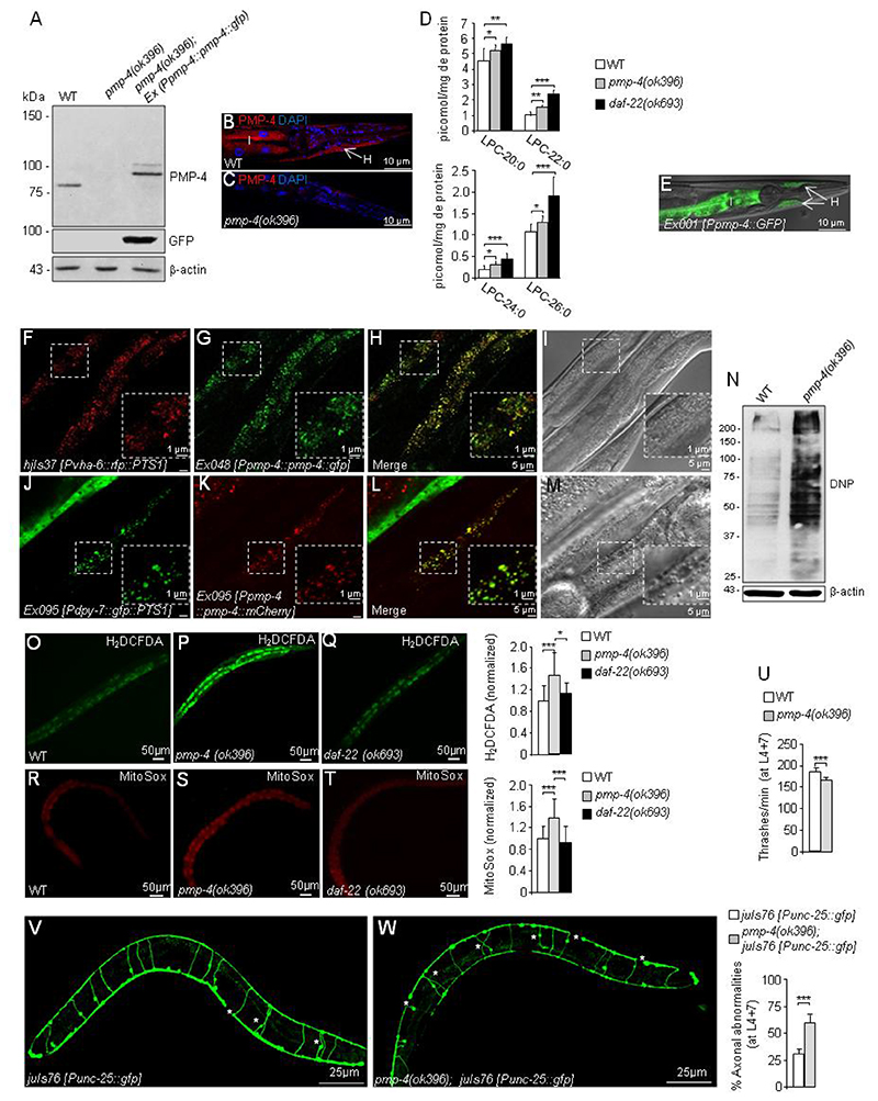
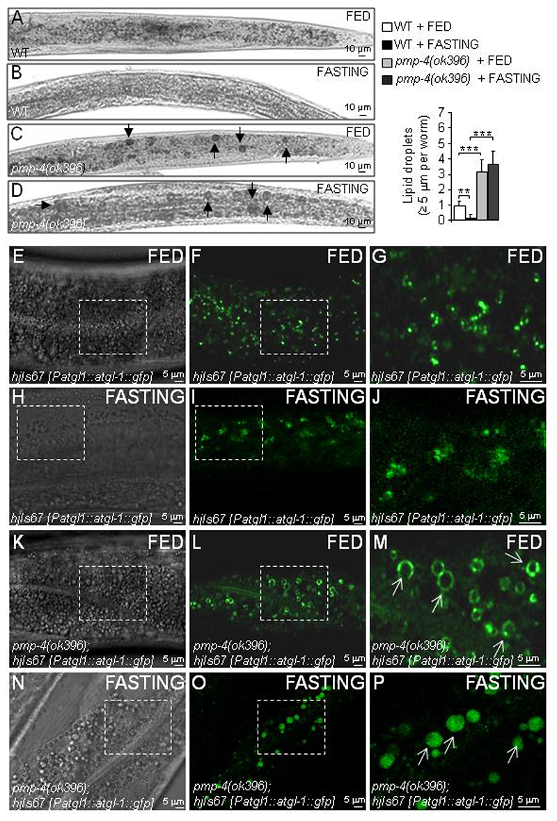
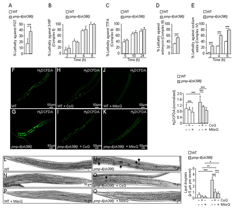
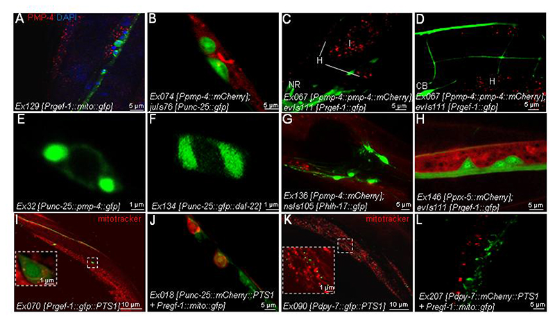
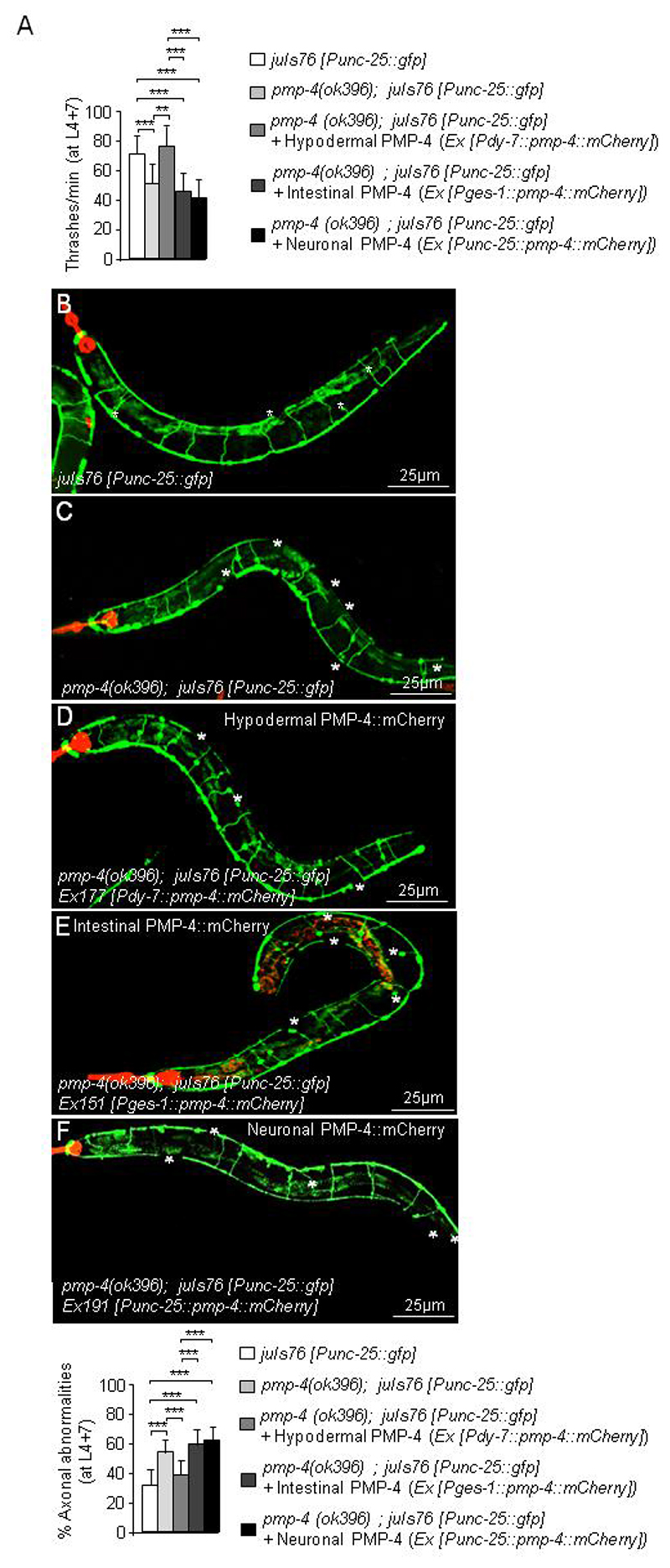
Adrenoleukodystrophy is a neurometabolic disorder caused by a defective peroxisomal ABCD1 transporter of very long-chain fatty acids (VLCFAs). Its pathogenesis is incompletely understood. Here we characterize a nematode model of X-ALD with loss of the pmp-4 gene, the worm orthologue of ABCD1. These mutants recapitulate the hallmarks of X-ALD: i) VLCFAs accumulation and impaired mitochondrial redox homeostasis and ii) axonal damage coupled to locomotor dysfunction. Furthermore, we identify a novel role for PMP-4 in modulating lipid droplet dynamics. Importantly, we show that the mitochondria targeted antioxidant MitoQ normalizes lipid droplets size, and prevents axonal degeneration and locomotor disability, highlighting its therapeutic potential. Moreover, PMP-4 acting solely in the hypodermis rescues axonal and locomotion abnormalities, suggesting a myelin-like role for the hypodermis in providing essential peroxisomal functions for the nematode nervous system.

Keywords: Axonal degeneration; Hypodermis; Lipid droplets; Mitochondria redox imbalance; Peroxisomes; X-linked adrenoleukodystrophy.


Conflict of interest statement

Declaration of competing interest The authors declare no conflict of interest.

Figures

Figure 1
Figure 1. C. elegans pmp-4(ok396) mutants recapitulate the main hallmarks observed in X-ALD.
(A) PMP-4 and PMP-4::GFP protein levels in wild-type (WT), pmp-4(ok396) and pmp-4(ok396) animals expressing PMP-4::GFP under the control of the pmp-4 promoter (pmp-4(ok396); Ex042 [Ppmp-4::pmp-4::gfp]) at the L4 larval stage. β-actin was used as a loading control (bottom panel) (n=4 pools of worms by condition). (B-C) Immunofluorescence staining of formaldehyde-fixed (B) WT and (C) pmp-4(ok396) worms incubated with polyclonal anti-PMP-4 (red) and counterstained with DAPI (blue) at the L4 larval stage. The posterior part of the worm is depicted. I=intestine and H=hypodermis. Scale = 10μm. (D) Lysophosphatidylcholine fatty acid levels (LPC-20:0, LPC-22:0, LPC-24:0 and LPC-26:0) of L4 worm lysates of WT (n=12), pmp-4(ok396)(n=10) and daf-22(ok693) animals (n=6). The daf-22(ok693) mutant was used as a positive control. (E) Tissular expression of GFP under the control of the pmp-4 promoter (Ex001 [Ppmp-4::gfp]) in the intestine=I and hypodermis=H at L4. The worm is oriented head to the left, dorsal face-up. The same results were obtained by three independent transgenic lines. Scale = 10μm. (F-I) The construct containing PMP-4::GFP under the control of the pmp-4 promoter was injected into the strain labelling peroxisomes with RFP in the intestine (hjIs37 [Pvha-6p::rfp::PTS1]; Ex048 [Ppmp-4::pmp-4::gfp]). Images of (F) RFP::PTS1 and (G) PMP-4::GFP staining. (H) Merge of images F and G. In yellow, colocalization of RFP::PTS-1 with PMP-4::GFP. (I) DIC-Nomarsky pictures corresponding to the fluorescence images (n=30). (J-M) The constructs containing GFP::PTS1 under the control of a specific hypodermal promoter (Pdpy-7) and PMP-4::mCherry under the control of the pmp-4 promoter were coinjected in the N2 strain (Ex095 [Pdpy-7::gfp::PTS1 + Ppmp-4::pmp-4::mCherry]). Here, the peroxisomes are labelled with GFP in the hypodermis. Images of (J) GFP::PTS1 and (K) PMP-4::mCherry staining. (L) Merge of images J and K. In yellow, colocalization of GFP::PTS-1 with PMP-4::mCherry. (M) DIC-Nomarsky images corresponding to the fluorescence images (n=30). Scale = 5 μm in each figure except for the expanded pictures in the squares, in which scale = 1 μm. The worms in the figures are oriented head to the left, dorsal face-up and only a portion of the central part of the body is depicted. (N) Dinitrophenol (DNP) protein levels in WT and pmp-4(ok396) animals at the L4 larval stage. The quantification of these blots by densitometry was performed and normalized to β-actin (n=4 pools of worms by condition). Relative total ROS levels (H2DCFDA) were measured by quantifying the fluorescence emission of the H2DCFDA probes in living animals in (O) WT (n=51), (P) pmp-4(ok396)(n=66) and (Q) daf-22(ok693) (n=40) L4 nematodes maintained in a liquid medium. Values are normalized to WT. Relative mitochondrial ROS levels in living animals in (R) WT (n=66), (S) pmp-4(ok396) (n=88) and (T) daf-22(ok693) (n=66) L4 nematodes quantified with MitoSox probes. Values are normalized to WT worms. Scale = 50 μm. (U) The thrashing behaviour of WT and pmp-4(ok396) nematodes was analysed in liquid medium at L4+7 days (n=20 animals/condition). Representative fluorescence images of GFP-labelled GABAergic neurons showing axonal abnormalities and quantitative analysis in living worms in (V) juIs76 [Punc-25::gfp] (n=20) and (W) pmp-4(ok396); juIs76 [Punc-25::gfp] (n=20) at L4+7 days. Scale = 25 μm. Data represent the mean ± standard deviation (SD). Statistical analysis was carried out with one-way ANOVA, followed by Tukey’s post hoc test (*P<0.05; **P<0.01; ***P<0.001) for D, O-S. Statistical analysis was carried out with Student’s t-test (*P<0.05; **P<0.01; ***P<0.001) for U-W.
Figure 2
Figure 2. pmp-4(ok396) worms display lipid droplet accumulation.
(A-P) At the L4 larval stage, WT and pmp-4(ok396) nematodes were fed or fasted for 24 h. (A-D) Bright field images of the posterior part of the worm stained with Sudan Black in (A) WT + FED (n=46), (B) WT + FASTING (n=60), (C) pmp-4(ok396) + FED (n=60), and (D) pmp-4(ok396) + FASTING (n=66). Lipids are evident as black droplets, labelled by a black arrow. Scale = 10 μm. Semi-quantitative analysis of the lipid droplets with a diameter >= 5 μm under the indicated conditions. (E-P) Localization of ATGL-1::GFP protein, driven by the atgl-1 promoter at L4, in (E-G) hjIs67 [Patgl-1::atgl-1::gfp] + FED, (H-J) hjIs67 [Patgl-1::atgl-1::gfp] + FASTING, (K-M) pmp-4(ok396); hjIs67 [Patgl-1::atgl-1::gfp] + FED and (N-P) pmp-4(ok396); hjIs67 [Patgl-1::atgl-1::gfp] + FASTING animals. (E) DIC-Nomarsky image and (F-G) GFP staining in WT. G is an enlarged image of F. (H) DIC-Nomarsky image and (I-J) GFP staining in WT after 24 h of fasting. J is an enlarged image of I. (K) DIC-Nomarsky image and (L-M) GFP staining in pmp-4(ok396). M is an enlarged image of L. (N) DIC-Nomarsky image and (O-P) GFP staining in pmp-4(ok396); hjIs67 [Patgl-1::atgl-1::gfp] after 24 h of fasting. P is an enlarged image of O. ATGL-1::GFP protein localized to the surface of large lipid droplets are indicated with white arrows (n=30 by conditions). The worms in the figures are oriented head to the left, dorsal face-up and only a portion of the central part of the body is depicted. Scale = 5 μM. Data represent the mean ± SD. Statistical analysis was carried out with two-way ANOVA, followed by Tukey’s post hoc test (*P<0.05; **P<0.01; ***P<0.001).
Figure 3
Figure 3. Mitochondrial dysfunction in pmp-4(ok396) animals.
WT and pmp-4(ok396) worms were incubated at the L4 larval stage with (A) the mitochondrial complex I inhibitor Paraquat (PQT) (n=51 to 54 animals by condition), the mitochondrial complex II inhibitors (B) 3-nitropropionic acid (3-NP) (n=15 to 20 animals by condition), (C) thenoyltrifluoroacetone (TTFA) (n=18 to 20 animals by condition), (D) the complex III inhibitor (antimycin A) (n=20 animals by condition) and (E) the complex IV inhibitor (sodium azide)(n=20 animals by condition). The lethality of the worms after the treatment was evaluated as described in the methodology. (F-Q). At L4, WT and pmp-4(ok396) nematodes were treated with CoQ (1 mg/ml) or MitoQ (5 μg/ml) for 7 days. Total ROS levels were measured by quantifying the fluorescence emission of the H2DCFDA probes in living animals in (F) WT (n=42), (G) pmp-4(ok396)(n=35), (H) WT + CoQ (n=32), (I) pmp-4(ok396) + CoQ (n=30), (J) WT + MitoQ (n=47), and (K) pmp-4(ok396) + MitoQ (n=32) animals at L4+7 days. Values are normalized to the untreated WT nematodes. Scale = 50 μm. Bright field images of the posterior part of the worm stained with Sudan Black in (L) WT (n=46), (M) pmp-4(ok396)(n=61), (N) WT + CoQ (n=60), (O) pmp-4(ok396) + CoQ (n=60), (P) WT + MitoQ (n=30), and (Q) pmp-4(ok396) + MitoQ (n=27) worms at L4+7 days. Lipids are evident as black droplets, labelled by a black arrow. Scale = 10 μm. Semi-quantitative analysis of the lipid droplets with a diameter >= 5 μm under the indicated conditions. Data represent the mean ± SD. Statistical analysis was carried out with Student’s t-test (*P<0.05; **P<0.01; ***P<0.001) for A-E. Statistical analysis was carried out with two-way ANOVA, followed by Tukey’s post hoc test (*P<0.05; **P<0.01; ***P<0.001) for F-Q.
Figure 4
Figure 4. Mitochondrial-targeted antioxidant rescue axonal abnormalities and locomotor dysfunction at old stages of development.
(A-G) At the L4 larval stages, WT and pmp-4(ok396) nematodes were treated with CoQ (1 mg/ml) or MitoQ (5 μg/ml) for 7 days. (A) The thrashing behaviour was carried out in WT and pmp-4(ok396) nematodes upon CoQ or MitoQ at L4+7 days (n=15 to 27 animals by condition). Representative fluorescence images showing axonal abnormalities and quantitative analysis at the indicated genotypes upon CoQ or MitoQ at L4+7 days in (B) juIs76 [Punc-25::gfp] (n=20), (C) pmp-4(ok396); juIs76 [Punc-25::gfp] (n=20), (D) juIs76 [Punc-25::gfp] + CoQ (n=18), (E)pmp-4(ok396); juIs76 [Punc-25::gfp] + CoQ (n=15), (F) juIs76 [Punc-25::gfp] + MitoQ (n=26) and (G) pmp-4(ok396; juIs76 [Punc-25::gfp]) + MitoQ (n=27). White asterisks indicate axonal abnormalities. All worms are oriented with the anterior end left and ventral side down. Scale = 25 μm. (H-N) At the L4 larval stage, juIs76 [Punc-25::gfp]and pmp-4(ok396); juIs76 [Punc-25::gfp] nematodes were treated with paraquat (PQT) (0.2 mM) and/or MitoQ (5 μg/ml) for 7 days. (H) The thrashing behaviour was analysed at stage L4+7 days in WT and pmp-4(ok396) nematodes upon PQT and/or MitoQ (n=20 to 31 animals by condition). Representative fluorescence images showing axonal abnormalities and quantitative analysis at stage L4+7 days in (I) juIs76 [Punc-25::gfp]) (n=29), (J) pmp-4(ok396); juIs76 [Punc-25::gfp] (n=31), (K) juIs76 [Punc-25::gfp] + PQT(n=25), (L) pmp-4(ok396); juIs76 [Punc-25::gfp] + PQT (n=20), (M) juIs76 [Punc-25::gfp] + PQT + MitoQ (n=21), (N) pmp-4(ok396; juIs76 [Punc-25::gfp] + PQT + MitoQ (n=28). Scale = 25μm. All worms are oriented with the anterior end left and ventral side down. Data represent the mean ± standard deviation (SD). Statistical analysis was carried out with two-way ANOVA, followed by Tukey’s post hoc test (*P<0.05; **P<0.01; ***P<0.001) for A-N.
Figure 5
Figure 5. Subcellular localization and tissue distribution of PMP-4
(A) mito::GFP was expressed under the control of the pan-neuronal promoter rgef-1 (Prgef-1) in WT animals (Ex129 [Prgef-1::mito::gfp]). Here, mitochondria located in all neurons are labelled with GFP. Next, worms were fixed and double immunostained against PMP-4 (red). Nuclei were counterstained with DAPI (blue) (n=30). Scale = 5 μm. (B) mCherry driven by the pmp-4 promoter was expressed in the GABAergic strain juIs76 [Punc-25::gfp], where GABAergic neurons are stained with GFP (juIs76 [Punc-25::gfp]; Ex074 [Ppmp-4::mCherry])(n=30). Scale = 5 μm. (C-D) PMP-4::mCherry driven by the pmp-4 promoter was expressed in a pan-neuronal strain where all neurons are stained with GFP (evIs111 [Prgef-1::gfp]; Ex067 [Ppmp-4::pmp-4::mCherry]) (n=30). Next, PMP-4::mCherry and GFP staining was observed in nematodes. Cell bodies (CB), NR: Nerve ring, H: hypodermis, I: intestine. Scale = 5μm. (E) PMP-4::GFP (Ex132 [Punc-25::pmp-4::gfp]) and (F) GFP::DAF-22 (Ex134 [Punc-25::gfp::daf-22]) under the control of a GABAergic promoter (Punc-25) were expressed in WT worms (n=30 by condition). (E) PMP-4::GFP and (F) GFP::DAF-22 staining was observed in nematodes. Scale = 1 μm. (G) mCherry driven by the pmp-4 promoter was expressed in glial-like cells strain nsIs105 [Phlh-17::gfp], where glial CEPsh cells are labelled with GFP (nsIs105 [Phlh-17::gfp]; Ex 136 [Ppmp-4::mCherry]) (n=30). Next, mCherry and GFP staining was observed in nematodes. Scale = 5 μm. (H) mCherry driven by the peroxisomal prx-5 promoter (Pprx-5) was expressed in the pan-neuronal strain evIs111 [Prgef-1::gfp], where all neurons are labelled with GFP (evIs111 [Prgef-1::gfp]; Ex146 [Pprx-5::mCherry]) (n=30). Next, mCherry and GFP staining were observed in nematodes. Scale = 5μm. We then used a construct containing PTS1 to target the peroxisomes. (I) GFP::PTS1 driven by a pan-neuronal promoter (Prgef-1) was expressed in WT worms (Ex070 [Prgef-1::gfp::PTS1]). Next, mitochondria were stained with Mitotracker probes, and GFP staining was visualized (n=30). Scale = 10 μm. (J) mCherry::PTS1 driven by a GABAergic promoter (Punc-25) and mito::GFP under the pan-neuronal promoter rgef-1 (Prgef-1) were co-expressed in WT animals (Ex218 [Punc-25::mCherry::PTS1 + Prgef-1::mito::gfp]). Next, mCherry and GFP staining were observed in nematodes (n=30). Scale = 5μm. (K) GFP::PTS1 driven by a hypodermal promoter (Pdpy-7) was expressed in WT worms (Ex090 [Pdpy-7::gfp::PTS1]). Next, mitochondria were stained with Mitotracker probes, and GFP staining was visualized (n=30). Scale = 10 μm. (L) mCherry::PTS1 driven by a hypodermal promoter (Pdpy-7) and mito::GFP under the pan-neuronal promoter rgef-1 (Prgef-1) were co-expressed in WT animals (Ex207 [Pdpy-7::mCherry::PTS1 + Prgef-1::mito::gfp]). Next, mCherry and GFP staining was observed in nematodes (n=30). Scale = 5 μm.
Figure 6
Figure 6. Specific PMP-4 expression in the hypodermis is essential to maintain axonal integrity and locomotion.
(A) Locomotion behaviour expressed as thrashes per minute at L4+7 days in juIs76 [Punc-25::gfp], pmp-4(ok396); juIs76 [Punc-25::gfp] and pmp-4(ok396; juIs76 [Punc-25::gfp]) animals where PMP-4::mCherry is expressed in hypodermis (pmp-4(ok396); juIs76 [Punc-25::gfp]; Ex177 [Pdpy-7::pmp-4::mCherry]), in intestine (pmp-4(ok396); juIs76 [Punc-25::gfp]; Ex151 [Pges-1::pmp-4::mCherry]) or in neurons (pmp-4(ok396); juIs76 [Punc-25::gfp]; Ex191 [Punc-25::pmp-4::mCherry]) (n= 19 to 23 animals by condition). Representative confocal pictures showing axonal damage and quantitative analysis in (B) juIs76 [Punc-25::gfp] (n=22), (C) pmp-4(ok396); juIs76 [Punc-25::gfp] animals (n=19), pmp-4(ok396) animals where PMP-4::mCherry is expressed in (D) hypodermis (pmp-4(ok396); juIs76 [Punc-25::gfp]; Ex177 [Pdpy-7::pmp-4::mCherry]) (n=23), (E) intestine (pmp-4(ok396); juIs76 [Punc-25::gfp]; Ex151 [Pges-l::pmp-4::mCherry]) (n=22) or (F) neurons (pmp-4(ok396); juIs76 [Punc-25::gfp]; Ex191 [Punc-25::pmp-4::mCherry]) (n=19). The red colour in all pictures corresponds to the strong expression in the pharynx of the co-injection marker Pmyo-2::mCherry. White asterisks label the axonal abnormalities. Worms are oriented head to the left and dorsal face-up. Scale = 25 μm. Data represent the mean ± standard deviation (SD). Statistical analysis was carried out with two-way ANOVA, followed by Tukey’s post hoc test (*P<0.05; **P<0.01; ***P<0.001).

Similar articles

Cited by

References

    1. Van Veldhoven PP, Baes M. Peroxisome deficient invertebrate and vertebrate animal models. Front Physiol. 2013;4:335. - PMC - PubMed
    1. Waterham HR, Ferdinandusse S, Wanders RJ. Human disorders of peroxisome metabolism and biogenesis. Biochim Biophys Acta. 2016;1863(5):922–33. - PubMed
    1. Moser AB, Fatemi A. Newborn Screening and Emerging Therapies for X-Linked Adrenoleukodystrophy. JAMA Neurol. 2018:e - PubMed
    1. van Roermund CW, Visser WF, Ijlst L, van Cruchten A, Boek M, Kulik W, Waterham HR, Wanders RJ. The human peroxisomal ABC half transporter ALDP functions as a homodimer and accepts acyl-CoA esters. Faseb J. 2008;22(12):4201–8. - PubMed
    1. Engelen M, Kemp S, Poll-The BT. X-linked adrenoleukodystrophy: pathogenesis and treatment. Curr Neurol Neurosci Rep. 2014;14(10):486. - PubMed

Publication types

MeSH terms

Substances