. 2020 Feb 4;12(3):2142-2155.
doi: 10.18632/aging.102731. Epub 2020 Feb 4.

Baicalin alleviates benign prostate hyperplasia through androgen-dependent apoptosis

Affiliations

Baicalin alleviates benign prostate hyperplasia through androgen-dependent apoptosis

Bo-Ram Jin et al. Aging (Albany NY). .

Abstract

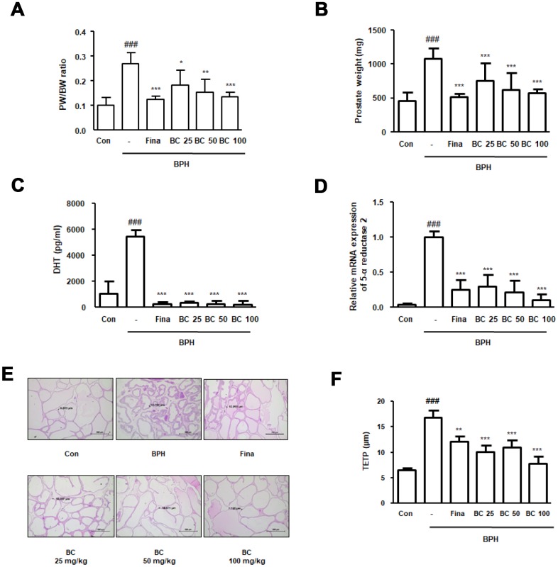
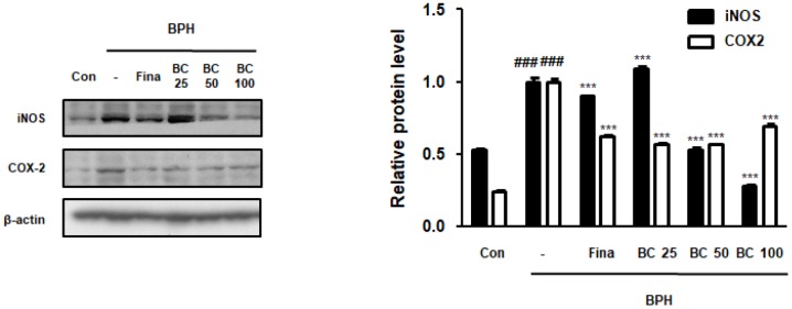
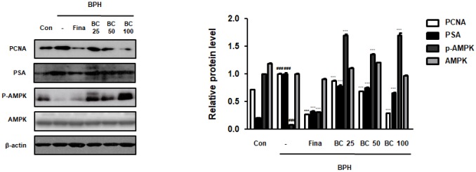
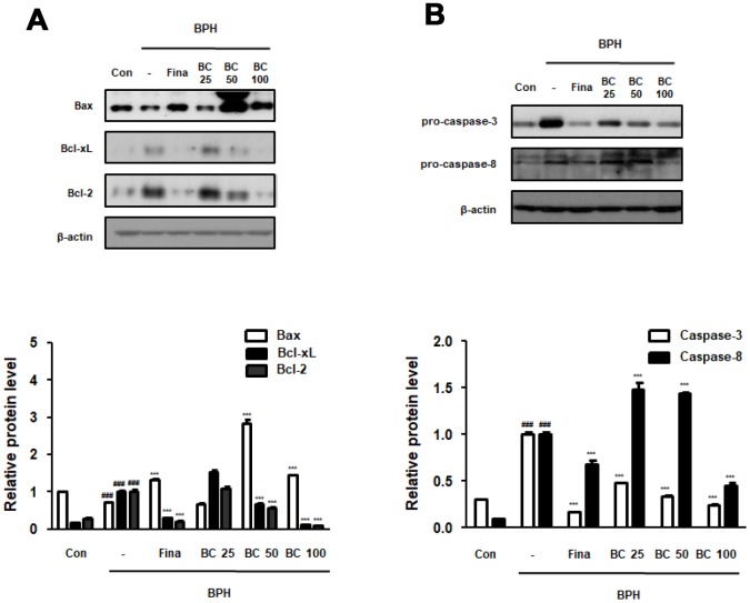
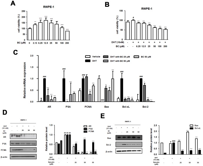
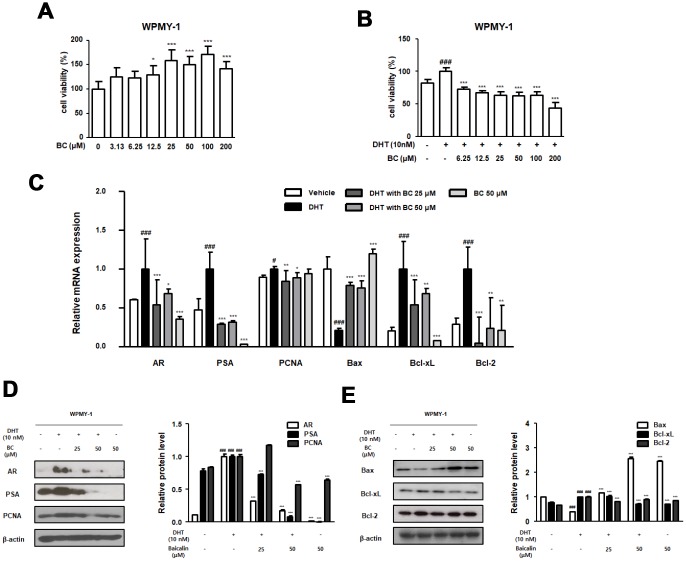
BPH is a disease prevalent among elderly men that is characterized by abnormal proliferation of prostatic epithelial and stromal tissues. No effective treatment exists for BPH owing to lack of a clear understanding of its molecular etiology. Although several studies have reported therapeutic effects of baicalin against numerous diseases, including prostate cancer, its beneficial effects on BPH have not yet been explored. The present study investigated the therapeutic effects of baicalin on the development of BPH and its mechanism of action. We established a testosterone-treated BPH animal model and DHT-stimulated prostate cell lines, including RWPE-1 and WPMY-1. Administration of baicalin ameliorated the pathological prostate enlargement, suppressed the production of DHT, and inhibited the activity of 5α- reductase Type II in the animal model. BC exerted these effects via its anti-proliferative effects by restoring the Bax/Bcl-2 ratio, activating caspase-3 and caspase-8, and inducing the phosphorylation of AMPK. In vitro studies using DHT-stimulated prostate cells demonstrated an up-regulation of BPH-related and proliferation markers, whereas baicalin clearly reduced the overexpression of AR, PSA, PCNA, and Bcl-2. These results suggested that baicalin could suppress androgen-dependent development of BPH both in vivo and in vitro by inducing apoptosis.

Keywords: androgen; apoptosis; baicalin; benign prostatic hyperplasia; proliferation.


Conflict of interest statement

CONFLICTS OF INTEREST: The authors declare no competing financial and non-financial interests.

Figures

Figure 1
Figure 1
Effect of BC on prostatic enlargement by inhibition of 5α-reductase Type II in TP-treated BPH rat model. Animals were injected with TP for 4 weeks with or without Fina and various concentration of BC. (A) Prostate weight to body weight (PW/BW) ratio and (B) prostate weight in each group were assessed. PW/BW ratio was calculated as (average prostate weight of the experimental group/ average body weight of the experimental group) × 1000. (C) DHT serum concentrations were analyzed using ELISA kit. (D) 5α-reductase Type II mRNA expression was showed by qRT-PCR analysis in prostatic tissues. (E) H&E staining analysis was performed using prostatic tissue sections. Original magnification 100x. (F) Based on H&E staining, TETP was measured and represented. All data are shown as the average value of each experimental group and are mean ± SD (n = 8). P value ### = P < 0.001 versus Con group; * = P < 0.05, ** = P <0.01, *** = P < 0.001 versus the BPH group.
Figure 2
Figure 2
Effect of BC on inflammatory makers in TP-treated BPH rats model. Prostatic tissue lysates were immunoblotted with iNOS and COX-2 to investigate effect of BC on compensatory cellular proliferation in TP-treated BPH rat model. β-actin served as an internal control. Fold changes in densitometric analysis was normalized to β-actin and are represented as mean ± SD, which are acquired Image J. P value ### = P < 0.001 versus Con group; *** = P < 0.001 versus the BPH group.
Figure 3
Figure 3
Effect of BC on the cell proliferation in TP-treated BPH rats model. Immunoblot results showed the level of PCNA, PSA, p-AMPK, AMPK in prostatic tissues. Densitometric protein levels of PCNA, PSA, p-AMPK, AMPK are represented as mean ± SD and plots of each protein were showed. P value ### = P < 0.001 versus Con group; *** = P < 0.001 versus the BPH group.
Figure 4
Figure 4
Effect of BC on apoptosis regulatory proteins in TP-treated BPH rats model. (A) The protein levels of Bcl-2 gene family and (B) pro-caspase-3 and pro-caspase-8 were determined via western blot using specific antibodies in prostatic tissue lysates. β-actin served as an internal control. Densitometric analysis on each protein was performed and relative protein levels were represented as mean ± SD. P value ### = P < 0.001 versus Con group; *** = P < 0.001 versus the BPH group.
Figure 5
Figure 5
Effect of BC on the BPH-related and apoptosis related protein and mRNA expression in RWPE-1 cells. (A) Effect of BC on the cell viability in RWPE-1 cells. RWPE-1 cells were treated with 3.13 to 200 μM of BC for 24 h. (B) Inhibitory effect of BC on cell proliferation in DHT-stimulated RWPE-1 cells. RWPE-1 cells were treated with 10 nM DHT, with or without 6.25–200 μM of BC for 24h. (CE) RWPE-1 were stimulated with 10 nM DHT from 3days to 5days, with or without BC (25, 50 μM). (C) The mRNA level of AR, PSA, PCNA, Bax, Bcl-xL and Bcl-2 were quantificated using RT-PCR. RWPE-1 cell lysates were immunoblotted with (D) AR, PSA, PCNA (E) Bax, Bcl-2 primary antibodies. Protein levels which are normalized by internal control β-actin are represented as relative protein levels. P value ### = P < 0.001 versus vehicle group; * = P < 0.05, ** = P <0.01, *** = P < 0.001 versus the DHT-stimulated group.
Figure 6
Figure 6
Effect of BC on the BPH-related and apoptosis related protein and mRNA expression in WPMY-1 cells. (A) Effect of BC on the cell viability in WPMY-1 cells. WPMY-1 cells were treated with 3.13 to 200 μM of BC for 24 h. (B) Inhibitory effect of BC on cell proliferation in DHT-stimulated WPMY-1 cells. The cells were treated with 10 nM DHT, with or without 6.25–200 μM of BC for 24h. (CE) WPMY-1 cells were stimulated with 10 nM DHT for 24h, with or without BC (25, 50 μM). (B) The mRNA level of AR, PSA, PCNA, Bax, Bcl-xL and Bcl-2 were quantificated using RT-PCR in DHT-stimulated WPMY-1 cells. Protein levels of (C) AR, PSA, PCNA (D) Bax, Bcl-xL and Bcl-2 are determined using western blotting in DHT-stimulated WPMY-1 cells. β-actin are used for internal control. Results are represented as mean ± SD. P value ### = P < 0.001 versus vehicle group; * = P < 0.05, ** = P <0.01, *** = P < 0.001 versus the DHT-stimulated group.

Similar articles

Cited by

References

    1. De Nunzio C, Aronson W, Freedland SJ, Giovannucci E, Parsons JK. The correlation between metabolic syndrome and prostatic diseases. Eur Urol. 2012; 61:560–70. 10.1016/j.eururo.2011.11.013 - DOI - PubMed
    1. Hammarsten J, Högstedt B, Holthuis N, Mellström D. Components of the metabolic syndrome-risk factors for the development of benign prostatic hyperplasia. Prostate Cancer Prostatic Dis. 1998; 1:157–62. 10.1038/sj.pcan.4500221 - DOI - PubMed
    1. Untergasser G, Madersbacher S, Berger P. Benign prostatic hyperplasia: age-related tissue-remodeling. Exp Gerontol. 2005; 40:121–28. 10.1016/j.exger.2004.12.008 - DOI - PubMed
    1. De Nunzio C, Kramer G, Marberger M, Montironi R, Nelson W, Schröder F, Sciarra A, Tubaro A. The controversial relationship between benign prostatic hyperplasia and prostate cancer: the role of inflammation. Eur Urol. 2011; 60:106–17. 10.1016/j.eururo.2011.03.055 - DOI - PubMed
    1. Lepor H. Pathophysiology, epidemiology, and natural history of benign prostatic hyperplasia. Rev Urol. 2004. (Suppl 9); 6:S3–10. - PMC - PubMed

Publication types

MeSH terms