. 2020 Jan 30;12(2):158.
doi: 10.3390/v12020158.

Diversity and Host Interactions Among Virulent and Temperate Baltic Sea Flavobacterium Phages

Affiliations

Diversity and Host Interactions Among Virulent and Temperate Baltic Sea Flavobacterium Phages

Emelie Nilsson et al. Viruses. .

Abstract

Viruses in aquatic environments play a key role in microbial population dynamics and nutrient cycling. In particular, bacteria of the phylum Bacteriodetes are known to participate in recycling algal blooms. Studies of phage-host interactions involving this phylum are hence important to understand the processes shaping bacterial and viral communities in the ocean as well as nutrient cycling. In this study, we isolated and sequenced three strains of flavobacteria-LMO6, LMO9, LMO8-and 38 virulent phages infecting them. These phages represent 15 species, occupying three novel genera. Additionally, one temperate phage was induced from LMO6 and was found to be competent at infecting LMO9. Functions could be predicted for a limited number of phage genes, mainly representing roles in DNA replication and virus particle formation. No metabolic genes were detected. While the phages isolated on LMO8 could infect all three bacterial strains, the LMO6 and LMO9 phages could not infect LMO8. Of the phages isolated on LMO9, several showed a host-derived reduced efficiency of plating on LMO6, potentially due to differences in DNA methyltransferase genes. Overall, these phage-host systems contribute novel genetic information to our sequence databases and present valuable tools for the study of both virulent and temperate phages.

Keywords: aquatic; genomics; host range; lysogeny; lytic.


Conflict of interest statement

The authors declare no conflict of interest.

Figures

Figure 1
Figure 1
Phylogenomic placement of the three strains from this study—LMO6, LMO8, and LMO9—in the GTDB phylogeny [71]. The three strains were placed with GTDBtk [70] and here visualised together with their closest Flavobacterium spp. relatives. Numbers at nodes are bootstrap values (values below 0.5 are not shown).
Figure 2
Figure 2
Similarity between the 38 lytic phages and the temperate phage based on Gegenees, which does a fragmented blastn between the entire genomes [67]; for details see Supplementary Table S3. Species are boxed with black while genera are coloured in the labels. Phages belonging to Muminvirus are orange, the isolate that constitutes Labanvirus is blue, Tantvirus is green and the isolates that belong to Pippivirus are pink. The species are named with the Flavobacterium virus and then 1 = Snusmum, 2 = Hattifnatt, 3 = Stinky, 4 = Hemulen, 5 = Snork, 6 = Sniff, 7 = Lillamy, 8 = Morran, 9 = Filifjonk, 10 = Tooticki, 11 = Mymlan, 12 = Mumin, 13 = Laban, 14 = Tant, 15 = Pippi, 16 = Lotta.
Figure 3
Figure 3
VICTOR tree produced with FastME based on nucleotide identity calculated with the D0-formula [73], with the newly isolated phages and reference genomes that share gene similarity to the novel phages presented in this study (accession number in parenthesis). The phages from this study are coloured based on the genera, as described in Figure 2. Bootstrap values above 50 are shown.
Figure 4
Figure 4
Genomic organisation of the type phages for each genus described in this study. Figure created with EasyFig [85]; for detailed information regarding the genomes and genes, see Supplementary Tables S1, S4–S7. The genomes are displayed linearly for visualisation.
Figure 5
Figure 5
Coverage of viral reads (from well 103) against the bacterial contig that contained the prophage, laban6-1 (contig “NODE_12”).
Figure 6
Figure 6
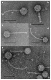
Negatively stained transmission electron micrographs of phage particles. (a) tant8-1, (b) pippi8-1, (c) mumin9-1, (d) laban6-1, (e) (i) tooticki6-1 and (e) (ii) laban6-1. Scale bars are 100 nm.
Figure 7
Figure 7
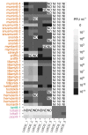
Host range tests for the type phages within each species with the three bacterial strains used within this study. ND = not determined, i.e., when the plaque assay has not been made; NI = no infection, i.e., when a plaque assay was performed but no plaques were detected. Phages are coloured according to genus, as in Figure 2. The number after, e.g., “mumin” indicates which bacterial strain the phage was originally isolated on, namely, 6 for LMO6, 8 for LMO8 and 9 for LMO9. The names on the x-axis are which bacterial strain the efficiency of plating was performed on, as well as if it was the first, second or third replicate (_1, _2, or _3).

Similar articles

Cited by

References

    1. Bergh O., Borsheim K.Y., Bratbak G., Heldal M. High abundance of viruses found in aquatic environments. Nature. 1989;340:467–468. doi: 10.1038/340467a0. - DOI - PubMed
    1. Suttle C.A. Marine viruses-major players in the global ecosystem. Nat. Rev. Microbiol. 2007;5:801–812. doi: 10.1038/nrmicro1750. - DOI - PubMed
    1. Weitz J.S., Poisot T., Meyer J.R., Flores C.O., Valverde S., Sullivan M.B., Hochberg M.E. Phage-bacteria infection networks. Trends Microbiol. 2013;21:82–91. doi: 10.1016/j.tim.2012.11.003. - DOI - PubMed
    1. Holmfeldt K., Solonenko N., Howard-Varona C., Moreno M., Malmstrom R.R., Blow M.J., Sullivan M.B. Large-scale maps of variable infection efficiencies in aquatic Bacteroidetes phage-host model systems. Environ. Microbiol. 2016;18:3949–3961. doi: 10.1111/1462-2920.13392. - DOI - PubMed
    1. Mann N.H., Cook A., Millard A., Bailey S., Clokie M. Marine ecosystems: Bacterial photosynthesis genes in a virus. Nature. 2003;424:741. doi: 10.1038/424741a. - DOI - PubMed

Publication types