

Dendritic Spines in Alzheimer's Disease: How the Actin Cytoskeleton Contributes to Synaptic Failure
- PMID: 32019166
- PMCID: PMC7036943
- DOI: 10.3390/ijms21030908
Dendritic Spines in Alzheimer's Disease: How the Actin Cytoskeleton Contributes to Synaptic Failure
Abstract
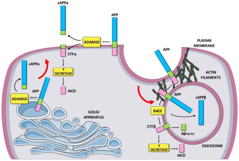
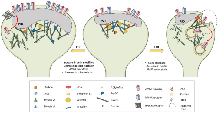
Alzheimer's disease (AD) is a neurodegenerative disorder characterized by Aβ-driven synaptic dysfunction in the early phases of pathogenesis. In the synaptic context, the actin cytoskeleton is a crucial element to maintain the dendritic spine architecture and to orchestrate the spine's morphology remodeling driven by synaptic activity. Indeed, spine shape and synaptic strength are strictly correlated and precisely governed during plasticity phenomena in order to convert short-term alterations of synaptic strength into long-lasting changes that are embedded in stable structural modification. These functional and structural modifications are considered the biological basis of learning and memory processes. In this review we discussed the existing evidence regarding the role of the spine actin cytoskeleton in AD synaptic failure. We revised the physiological function of the actin cytoskeleton in the spine shaping and the contribution of actin dynamics in the endocytosis mechanism. The internalization process is implicated in different aspects of AD since it controls both glutamate receptor membrane levels and amyloid generation. The detailed understanding of the mechanisms controlling the actin cytoskeleton in a unique biological context as the dendritic spine could pave the way to the development of innovative synapse-tailored therapeutic interventions and to the identification of novel biomarkers to monitor synaptic loss in AD.
Keywords: actin cytoskeleton; actin-binding proteins; amyloid; synaptic plasticity; synaptopathy.
Conflict of interest statement
The authors declare no conflict of interest. The funders had no role in the design of the study; in the collection, analyses, or interpretation of data; in the writing of the manuscript, or in the decision to publish the results.
Figures
References
-
- McKhann G.M., Knopman D.S., Chertkow H., Hyman B.T., Jack C.R., Kawas C.H., Klunk W.E., Koroshetz W.J., Manly J.J., Mayeux R., et al. The diagnosis of dementia due to Alzheimer’s disease: Recommendations from the National Institute on Aging-Alzheimer‘s Association workgroups on diagnostic guidelines for Alzheimer’s disease. Alzheimers Dement. 2011;7:263–269. doi: 10.1016/j.jalz.2011.03.005. - DOI - PMC - PubMed
-
- Vermunt L., Sikkes S.A.M., van den Hout A., Handels R., Bos I., van der Flier W.M., Kern S., Ousset P.-J., Maruff P., Skoog I., et al. Alzheimer Disease Neuroimaging Initiative; AIBL Research Group; ICTUS/DSA study groups Duration of preclinical, prodromal, and dementia stages of Alzheimer’s disease in relation to age, sex, and APOE genotype. Alzheimers Dement. 2019;15:888–898. doi: 10.1016/j.jalz.2019.04.001. - DOI - PMC - PubMed
