. 2020 Jan 30;21(3):900.
doi: 10.3390/ijms21030900.

A Unique Regulation Region in the 3' UTR of HLA-G with a Promising Potential

Affiliations

A Unique Regulation Region in the 3' UTR of HLA-G with a Promising Potential

Adi Reches et al. Int J Mol Sci. .

Abstract

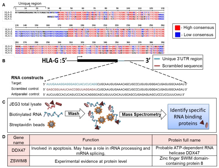
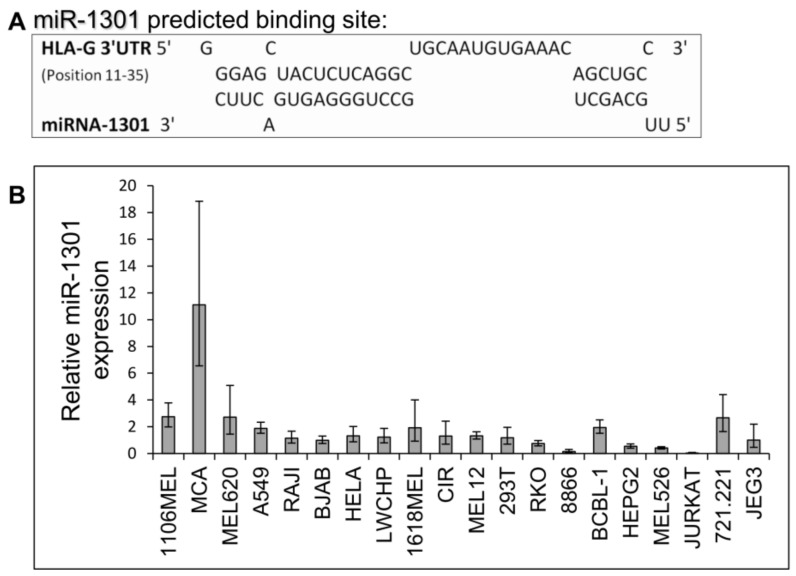
Human leukocyte antigen G (HLA-G) is a non-classical human leukocyte antigen (HLA) class I protein that interacts with inhibitory receptors and is commonly overexpressed in various cancers, thereby establishing itself as an inhibitory checkpoint immune ligand. It is also expressed in trophoblast cells during pregnancy and protects the fetus from immune rejection. Despite its crucial role and its intriguing expression pattern, the regulation of HLA-G's expression is only partially understood. HLA-G's mRNA is expressed in many tissues but the protein expression is restricted only to the cells mentioned above. Therefore, we suggest that HLA-G is post-transcriptionally regulated. Here, we reveal a distinctive site present only in the 3' Untranslated region (UTR) of HLA-G, which might explain its unique expression pattern. Consequently, we attempted to find binding factors such as RNA binding proteins (RBPs) and microRNAS (miRs) that regulate HLA-G expression by interacting with this distinct site present in its 3' UTR. Our research indicates that this site should be further studied in order to reveal its significance.

Keywords: 3′ UTR; HLA-G; RNA binding proteins; microRNA.


Conflict of interest statement

The authors declare that they have no conflict of interest.

Figures

Figure 1
Figure 1
Identification of RBPs that interacted with the unique part of HLA-G 3′ UTR. (A) Sequence alignment of the 3′ UTRs of HLA-G, -A, -B, and –C (Reference Sequences code respectively: NM_002127.5, LC257690.1, BC091497.1, MN848254.1). The alignment was performed using the MultAlin bioinformatics tool [25]. Identical nucleotides are indicated in red. Blue nucleotides represent similarities. (B) Schematic representation of the RNA constructs used in the RNA affinity purification assay. (C) Schematic representation of the RNA affinity purification assay. (D) Top two identified RBP candidates.
Figure 2
Figure 2
DDX47 does not influence HLA-G expression in cells expressing the HLA-G protein. (A,C,E) His-tagged DDX47 was overexpressed (OE) in JEG3 (A), 721.221 overexpressing HLA-G (C), JURKAT overexpressing HLA-G (E). Western blots were performed with anti-His-tag specific mAb and expression was compared to cells expressing an empty vector. Vinculin was used as a loading control. (B,D,F) Flow cytometry analysis of HLA-G (left) or classical HLA class I (right) expression on JEG3 (B), 721.221 overexpressing HLA-G (D), and JURKAT overexpressing HLA-G (F). Cells overexpressing His-tagged DDX47 or empty vector are represented by black and gray histograms, respectively. The filled gray histogram represents staining of cells with secondary mAb only. The figure shows one representative experiment out of three performed.
Figure 3
Figure 3
DDX47 does not induce HLA-G protein expression in cells expressing HLA-G at the mRNA level only. (A) HLA-G mRNA expression levels in various cell lines was evaluated using qRT-PCR. The figure shows fold enrichment of HLA-G in the indicated cell lines compared to hGAPDH. JEG3 cell line was set to 1. Shown are means ± Standard error of the mean (SEM) of triplicates. Figure shows one representative experiment out of three performed. (B,C,D) His-tagged DDX47 RBP was overexpressed (OE) in C1R (B), RAJI (C), and BJAB (D). Western blots were performed with anti-His-tag specific mAb and expression was compared with cells expressing empty vector. Vinculin was used as a loading control. (E,F,G) Flow cytometry analysis of HLA-G (upper panel) or classical HLA class I (lower panel) expression on C1R (E), RAJI (F), and BJAB (G) cells overexpressing His-tagged DDX47 or empty vector (black and gray histograms, respectively). The filled gray histogram represents staining of cells with secondary mAb only.
Figure 4
Figure 4
ZSWIM8 does not influence HLA-G expression. (A) KD of ZSWIM8 in JEG3 cells was performed using specific shRNAs (numbers indicate the shRNA catalog number). Western blots were performed with an anti-ZSWIM8 specific mAb. JEG3 cells expressing scrambled shRNA ZSWIM8 expression were set to 1. hGAPDH was used for loading control normalization. (B,C) Flow cytometry analysis of the expression of HLA-G (B) or classical HLA class I (C) on JEG3 cells transduced with the indicated shRNAs against ZSWIM8 (black histograms) compared to scrambled shRNA (gray histograms). The filled gray histogram represents the staining of scrambled shRNA with secondary mAb only. The background of the shRNA was similar to the scrambled shRNA and is not shown in the figure. Figures show one representative experiment out of three performed.
Figure 5
Figure 5
miR-1301 has a predicted binding site in the unique part of HLA-G 3′ UTR. (A) Predicted binding site between the unique part of HLA-G 3′ UTR (top) and hsa-miR-1301-3p (bottwm) using RNAhybrid bioinformatics tool [30]. (B) hsa-miR-1301-3p expression levels in various cell lines was evaluated using qRT-PCR. The figure shows fold enrichment of the miR in the indicated cell lines compared to hGAPDH. JEG3 cell line was set as 1. Shown are mean ± SEM of triplicates. Figure shows one representative experiment out of three performed.
Figure 6
Figure 6
miR-1301 does not influence HLA-G expression. (A,C) hsa-miR-1301-3p was overexpressed in JEG3 (A) and 721.221 overexpressing HLA-G (C). Expression was evaluated using qRT-PCR and compared to cells expressing empty vector (EV). (B,D) Flow cytometry analysis of HLA- (left) or classical HLA class I (right) expression on JEG3 (B) or 721.221 overexpressing HLA-G (D) cells, overexpressing hsa-miR-1301-3p or empty vector (black and gray histograms, respectively). The filled gray histogram represents staining of cells with secondary mAb only. Figure shows one representative experiment out of three performed.
Figure 7
Figure 7
Simultaneous expression of both DDX47 and miR-1301 does not influence HLA-G protein expression. (A,C) His-tagged DDX47 was overexpressed (OE) in JEG3 (A) and 721.221 overexpressing HLA-G (C). All the cells also overexpressed miR-1301 as verified in Figure 6A,C. Western blots were performed with anti-His-tag specific mAb and expression was compared to cells expressing empty vector. Vinculin was used as a loading control. (B,D) Flow cytometry analysis of HLA-G (left) or classical HLA class I (right) expression on JEG3 (B) and 721.221 overexpressing HLA-G (D) cells overexpressing His-tagged DDX47 or empty vector (black and gray histograms, respectively). The filled gray histogram represents staining of cells with secondary mAb only. Figure shows one representative experiment out of three performed.

References

    1. Niederkorn J.Y. Immune escape mechanisms of intraocular tumors. Prog. Retin. Eye Res. 2009;28:329–347. doi: 10.1016/j.preteyeres.2009.06.002. - DOI - PMC - PubMed
    1. Pratheek B.M., Nayak T.K., Sahoo S.S., Mohanty P.K., Chattopadhyay S., Chakraborty N.G., Chattopadhyay S. Mammalian non-classical major histocompatibility complex I and its receptors: Important contexts of gene, evolution, and immunity. Indian J. Hum. Genet. 2014;20:129–141. - PMC - PubMed
    1. Carosella E.D., Rouas-Freiss N., Roux D.T., LeMoreau P., Le Maoult J. Advances in Immunology. Volume 127. Academic Press Inc.; Cambridge, MA, USA: 2015. HLA-G. An Immune Checkpoint Molecule; pp. 33–144. - PubMed
    1. Clements C.S., Kjer-Nielsen L., McCluskey J., Rossjohn J. Structural Studies on HLA-G: Implications for Ligand and Receptor Binding. Hum. Immunol. 2007;68:220–226. doi: 10.1016/j.humimm.2006.09.003. - DOI - PubMed
    1. LeMaoult J., Zafaranloo K., Le Banff C., Carosella E.D. HLA-G up-regulates ILT2, ILT3, ILT4, and KIR2DL4 in antigen presenting cells, NK cells, and T cells. FASEB J. 2005;19:662–664. doi: 10.1096/fj.04-1617fje. - DOI - PubMed

MeSH terms