. 2020 Jan 30;21(3):917.
doi: 10.3390/ijms21030917.

Polyethylene Glycol 35 (PEG35) Protects against Inflammation in Experimental Acute Necrotizing Pancreatitis and Associated Lung Injury

Affiliations

Polyethylene Glycol 35 (PEG35) Protects against Inflammation in Experimental Acute Necrotizing Pancreatitis and Associated Lung Injury

Ana Ferrero-Andrés et al. Int J Mol Sci. .

Abstract

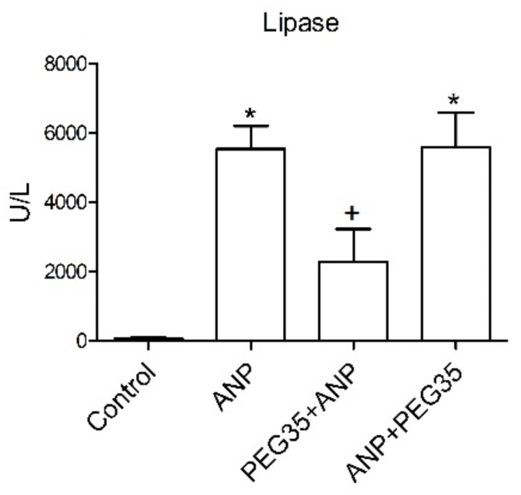
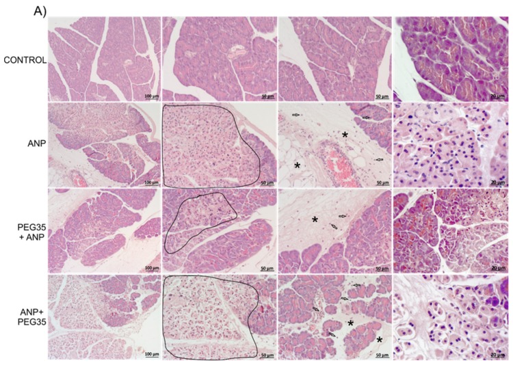
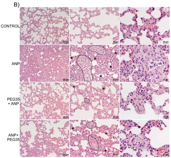
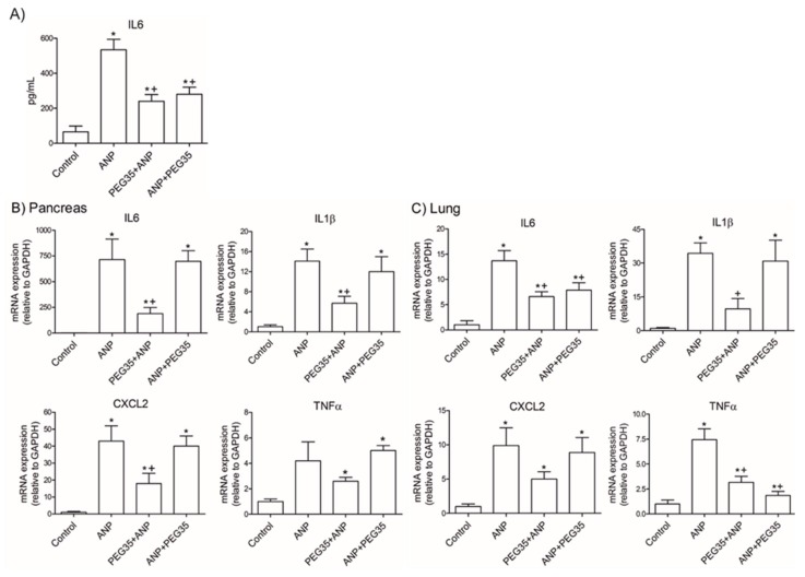
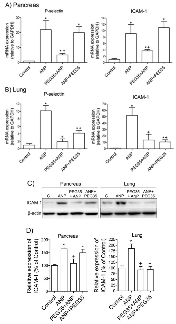
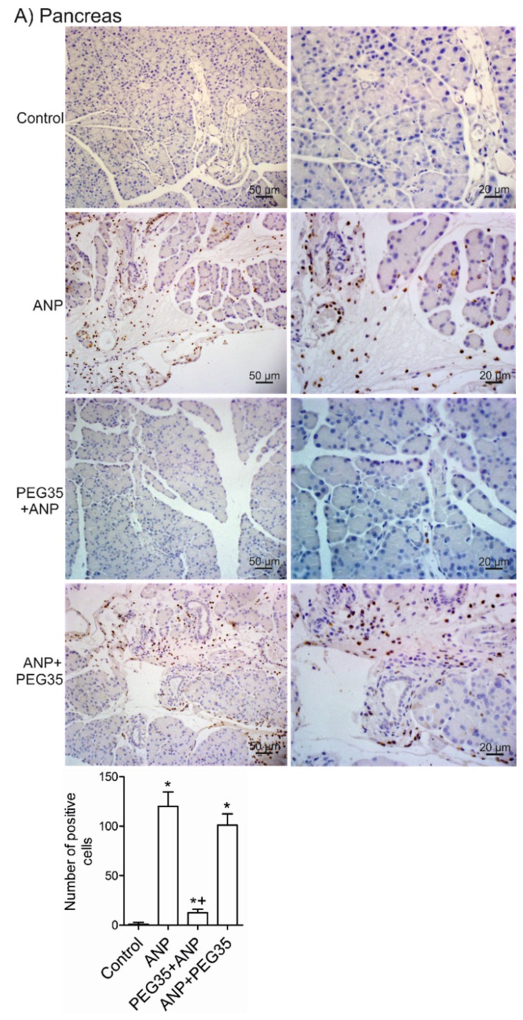
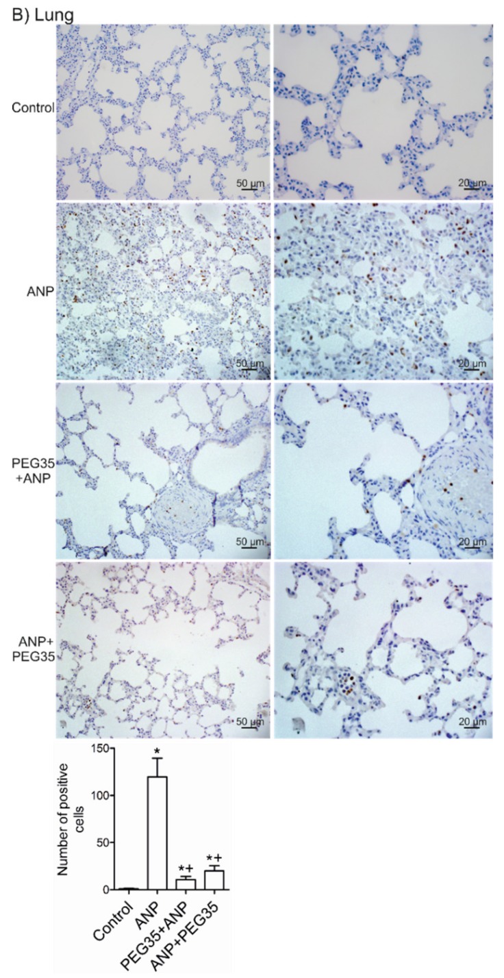
Acute pancreatitis is an inflammatory disorder of the pancreas. Its presentation ranges from self-limiting disease to acute necrotizing pancreatitis (ANP) with multiorgan failure and a high mortality. Polyethylene glycols (PEGs) are non-immunogenic, non-toxic, and water-soluble chemicals composed of repeating units of ethylene glycol. The present article explores the effect of PEG35 administration on reducing the severity of ANP and associated lung injury. ANP was induced by injection of 5% sodium taurocholate into the biliopancreatic duct. PEG35 was administered intravenously either prophylactically or therapeutically. Three hours after ANP induction, pancreas and lung tissue samples and blood were collected and ANP severity was assessed. To evaluate the inflammatory response, gene expression of pro-inflammatory cytokines and chemokine and the changes in the presence of myeloperoxidase and adhesion molecule levels were determined in both the pancreas and the lung. To evaluate cell death, lactate dehydrogenase (LDH) activity and apoptotic cleaved caspase-3 localization were determined in plasma and in both the pancreatic and lung tissue respectively. ANP-associated local and systemic inflammatory processes were reduced when PEG35 was administered prophylactically. PEG35 pre-treatment also protected against acute pancreatitis-associated cell death. Notably, the therapeutic administration of PEG35 significantly decreased associated lung injury, even when the pancreatic lesion was equivalent to that in the untreated ANP-induced group. Our results support a protective role of PEG35 against the ANP-associated inflammatory process and identify PEG35 as a promising tool for the treatment of the potentially lethal complications of the disease.

Keywords: cytokines; inflammation; pancreatitis; polyethylene glycols; pulmonary injury.


Conflict of interest statement

The authors declare no conflict of interest.

Figures

Figure 1
Figure 1
Effect of PEG35 treatment on plasma lipase activity in sodium taurocholate-induced ANP. Plasma lipase levels in U/L. Bars represent mean values of each group ± SEM. * p < 0.05 versus Control, + p < 0.05 versus ANP. ANP, Acute Necrotizing Pancreatitis. PEG35, Polyethylene glycol 35. Each determination was carried out in triplicate.
Figure 2
Figure 2
Effect of PEG35 treatment on histological changes in experimental acute necrotizing pancreatitis and associated acute lung injury. (A) Representative images of hematoxylin and eosin-stained pancreatic sections for each experimental group. Control group showed normal pancreas structure. ANP and ANP+PEG35 groups presented large areas of necrosis (under area), infiltrated polymorphonuclear neutrophils (indicated by empty arrows) and interstitial edema (indicated by an asterisk). Prophylactic administration of PEG35 significantly reduced these features. (B) Representative images of hematoxylin and eosin-stained lung sections for each experimental group. Control group showed normal alveolar structure. In the ANP group, a marked alveolar septal thickening (indicated by an asterisk) with infiltrated neutrophils, and the presence of vessel neutrophils (under area) were seen. Both prophylactic and therapeutic PEG35 treatment normalized alveolar septal thickening and neutrophils infiltration. ANP, Acute Necrotizing Pancreatitis. PEG35, Polyethylene glycol 35. Scale bar, 100, 50 and 20 µM.
Figure 2
Figure 2
Effect of PEG35 treatment on histological changes in experimental acute necrotizing pancreatitis and associated acute lung injury. (A) Representative images of hematoxylin and eosin-stained pancreatic sections for each experimental group. Control group showed normal pancreas structure. ANP and ANP+PEG35 groups presented large areas of necrosis (under area), infiltrated polymorphonuclear neutrophils (indicated by empty arrows) and interstitial edema (indicated by an asterisk). Prophylactic administration of PEG35 significantly reduced these features. (B) Representative images of hematoxylin and eosin-stained lung sections for each experimental group. Control group showed normal alveolar structure. In the ANP group, a marked alveolar septal thickening (indicated by an asterisk) with infiltrated neutrophils, and the presence of vessel neutrophils (under area) were seen. Both prophylactic and therapeutic PEG35 treatment normalized alveolar septal thickening and neutrophils infiltration. ANP, Acute Necrotizing Pancreatitis. PEG35, Polyethylene glycol 35. Scale bar, 100, 50 and 20 µM.
Figure 3
Figure 3
Role of PEG35 on the modulation of pro-inflammatory cytokines and chemokines expression in sodium taurocholate acute necrotizing pancreatitis and associated acute lung injury. (A) IL6 expression levels in plasma. (B) Pancreatic tissue gene expression of IL6, IL1β, CXCL2 and TNFα by real-time qRT-PCR. (C) Lung tissue gene expression of IL6, IL1β, CXCL2 and TNFα by real-time qRT-PCR. In all cases, mRNA induction levels were normalized to GAPDH mRNA expression. Bars represent mean values of each group ± SEM. * p < 0.05 versus Control, + p < 0.05 versus ANP. ANP, Acute Necrotizing Pancreatitis. PEG35, Polyethylene glycol 35. Each determination was carried out in triplicate.
Figure 4
Figure 4
Effects of PEG35 administration on the expression of adhesion molecules in acute necrotizing pancreatitis and associated acute lung injury. (A) Pancreatic tissue gene expression of P-selectin and ICAM-1 by real-time qRT-PCR. (B) Lung tissue gene expression of P-selectin and ICAM-1 by real-time qRT-PCR. In all cases, mRNA induction levels were normalized to GAPDH mRNA expression. (C) Pancreatic and pulmonar protein expression of ICAM-1 assessed by Western Blot analysis. Β-actin expression was used as loading control. Data shown are representative blots for each group. (D) Densitometry quantification of Western blot for ICAM-1 in pancreatic and lung tissue. Bars represent mean values of each group ± SEM. * p < 0.05 versus Control, + p < 0.05 versus ANP. C, Control. ANP, Acute Necrotizing Pancreatitis. PEG35, Polyethylene glycol 35. Each determination was carried out in triplicate.
Figure 5
Figure 5
Effects of PEG35 treatment on acute necrotizing pancreatitis-induced myeloperoxidase expression. (A) Top, Representative images of pancreatic sections stained with anti-MPO antibody (brown). Bottom, Pancreas MPO immunostaining quantification represented as the average number of positive cells per field. (B) Top, Representative images of lung sections stained with anti-MPO antibody (brown). Bottom, Pulmonary MPO immunostaining quantification represented as the average number of positive cells per field. ANP, Acute Necrotizing Pancreatitis. PEG35, Polyethylene glycol 35. Scale bar, 50 and 20 µM. * p < 0.05 versus Control, + p < 0.05 versus ANP.sss
Figure 5
Figure 5
Effects of PEG35 treatment on acute necrotizing pancreatitis-induced myeloperoxidase expression. (A) Top, Representative images of pancreatic sections stained with anti-MPO antibody (brown). Bottom, Pancreas MPO immunostaining quantification represented as the average number of positive cells per field. (B) Top, Representative images of lung sections stained with anti-MPO antibody (brown). Bottom, Pulmonary MPO immunostaining quantification represented as the average number of positive cells per field. ANP, Acute Necrotizing Pancreatitis. PEG35, Polyethylene glycol 35. Scale bar, 50 and 20 µM. * p < 0.05 versus Control, + p < 0.05 versus ANP.sss
Figure 6
Figure 6
Effects of PEG35 treatment on ANP-induced cell death. (A) Plasma lactate dehydrogenase activity three hours after acute necrotizing pancreatitis induction expressed as mU/mL. (B) Top: Representative images of pancreatic sections stained with anti-cleaved caspase-3 antibody (brown). Bottom: Pancreas cleaved caspase-3 immunostaining quantification represented as the percentage of marked surface per field. (C) Top: Representative images of lung sections stained with anti-cleaved caspase-3 antibody (brown). Bottom: Pulmonary cleaved caspase-3 immunostaining quantification represented as the average number of positive cells per field. Bars represent mean values of each group ± SEM. * p < 0.05 versus Control, + p < 0.05 versus ANP. ANP, Acute Necrotizing Pancreatitis. PEG35, Polyethylene glycol 35. Each determination was carried out in triplicate.

Similar articles

Cited by

References

    1. Lankisch P.G., Apte M., Banks P.A. Acute pancreatitis. Lancet. 2015;386:85–96. doi: 10.1016/S0140-6736(14)60649-8. - DOI - PubMed
    1. Banks P.A., Bollen T.L., Dervenis C., Gooszen H.G., Johnson C.D., Sarr M.G., Tsiotos G.G., Vege S.S. Acute Pancreatitis Classification Working G: Classification of acute pancreatitis—2012: Revision of the Atlanta classification and definitions by international consensus. Gut. 2013;62:102–111. doi: 10.1136/gutjnl-2012-302779. - DOI - PubMed
    1. Akbarshahi H., Rosendahl A.H., Westergren-Thorsson G., Andersson R. Acute lung injury in acute pancreatitis—Awaiting the big leap. Respir. Med. 2012;106:1199–1210. doi: 10.1016/j.rmed.2012.06.003. - DOI - PubMed
    1. Turecek P.L., Bossard M.J., Schoetens F., Ivens I.A. PEGylation of Biopharmaceuticals: A Review of Chemistry and Nonclinical Safety Information of Approved Drugs. J. Pharm. Sci. 2016;105:460–475. doi: 10.1016/j.xphs.2015.11.015. - DOI - PubMed
    1. Ivens I.A., Achanzar W., Baumann A., Brändli-Baiocco A., Cavagnaro J., Dempster M., Depelchin B.O., Rovira A.R., Dill-Morton L., Lane J.H., et al. PEGylated Biopharmaceuticals: Current Experience and Considerations for Nonclinical Development. Toxicol. Pathol. 2015;43:959–983. doi: 10.1177/0192623315591171. - DOI - PubMed

MeSH terms