. 2020 Jan 30;9(2):325.
doi: 10.3390/cells9020325.

Polyglutamine-Expanded Androgen Receptor Alteration of Skeletal Muscle Homeostasis and Myonuclear Aggregation Are Affected by Sex, Age and Muscle Metabolism

Affiliations

Polyglutamine-Expanded Androgen Receptor Alteration of Skeletal Muscle Homeostasis and Myonuclear Aggregation Are Affected by Sex, Age and Muscle Metabolism

Mathilde Chivet et al. Cells. .

Abstract

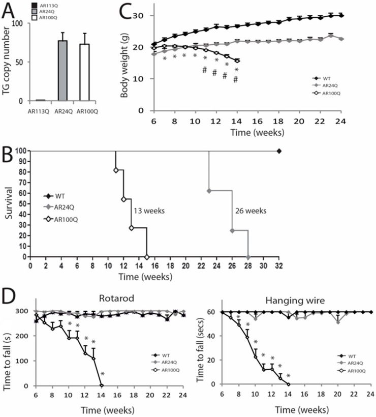
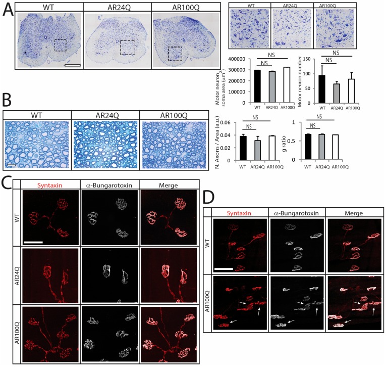
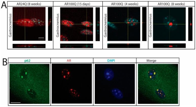
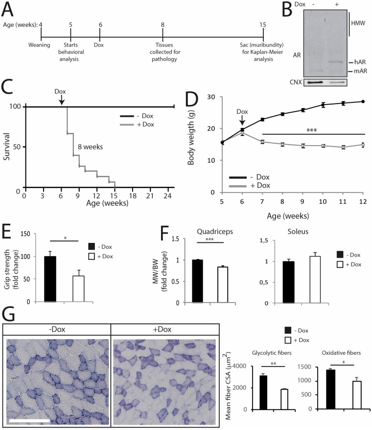
Polyglutamine (polyQ) expansions in the androgen receptor (AR) gene cause spinal and bulbar muscular atrophy (SBMA), a neuromuscular disease characterized by lower motor neuron (MN) loss and skeletal muscle atrophy, with an unknown mechanism. We generated new mouse models of SBMA for constitutive and inducible expression of mutant AR and performed biochemical, histological and functional analyses of phenotype. We show that polyQ-expanded AR causes motor dysfunction, premature death, IIb-to-IIa/IIx fiber-type change, glycolytic-to-oxidative fiber-type switching, upregulation of atrogenes and autophagy genes and mitochondrial dysfunction in skeletal muscle, together with signs of muscle denervation at late stage of disease. PolyQ expansions in the AR resulted in nuclear enrichment. Within the nucleus, mutant AR formed 2% sodium dodecyl sulfate (SDS)-resistant aggregates and inclusion bodies in myofibers, but not spinal cord and brainstem, in a process exacerbated by age and sex. Finally, we found that two-week induction of expression of polyQ-expanded AR in adult mice was sufficient to cause premature death, body weight loss and muscle atrophy, but not aggregation, metabolic alterations, motor coordination and fiber-type switch, indicating that expression of the disease protein in the adulthood is sufficient to recapitulate several, but not all SBMA manifestations in mice. These results imply that chronic expression of polyQ-expanded AR, i.e. during development and prepuberty, is key to induce the full SBMA muscle pathology observed in patients. Our data support a model whereby chronic expression of polyQ-expanded AR triggers muscle atrophy through toxic (neomorphic) gain of function mechanisms distinct from normal (hypermorphic) gain of function mechanisms.

Keywords: 2% SDS-resistant aggregates; androgen receptor; inclusion bodies; muscle metabolism; polyglutamine diseases; skeletal muscle.


Conflict of interest statement

The authors declare no conflict of interest.

Figures

Figure 1
Figure 1
AR100Q transgenic mice show reduced life span and progressive motor dysfunction. (A) Analysis of transgene copy number in knock-in (AR113Q) and transgenic (AR24Q and AR100Q) male mice (n = 5). (B) Kaplan-Meier survival curves of WT (n = 8), AR24Q (n = 8) and AR100Q (n = 11) male mice. Survival curves were compared using Log-rank (Mantel-Cox) test. (C) Temporal changes in mean BW of WT (n = 8), AR24Q (n = 7) and AR100Q (n = 14) male mice. (D) Temporal changes in mean rotarod and hanging wire task performance in WT (n = 7), AR24Q (n = 5) and AR100Q (n = 12) male mice. Graphs, mean ± SEM. Statistical testing: Mann Whitney test was used to test the difference between genotypes in (C) and (D). In (C) * p < 0.01 for AR100Q vs WT; # p < 0.001 for AR100Q vs AR24Q. In (D) * p < 0.05.
Figure 2
Figure 2
Late-onset alteration of NMJ morphology in AR100Q mice. (A) Nissl staining analysis of MN number and soma area in the lumbar spinal cord transversal sections of 12-week-old WT, AR24Q and AR100Q male mice (n = 3). Inset position is shown by the dashed square box. Bar, 500 micron. (B) Toluidine blue staining of semi-thin sciatic nerve transversal sections of 8-week-old WT, AR24Q and AR100Q male mice (n = 3). Bar, 10 micron. (CD) Immunohistochemical analysis of NMJ pathology in the quadriceps muscle of 8-week-old (C) and 12-week-old (D) WT, AR24Q and AR100Q male mice (n = 3). Arrows indicate fragmented NMJs. Bar, 50 microns. Shown are representative images. Graphs, mean ± SEM. Statistical testing: One-way ANOVA followed by Newman-Keuls post-hoc test; NS, non-significant.
Figure 3
Figure 3
Signs of muscle atrophy at the late stage of disease in AR100Q mice and not AR24Q mice. (A) Real-time PCR analysis in the quadriceps of WT, AR24Q and AR100Q male mice (n = 3–5). (B) H/E staining of transversal sections of quadriceps muscle of WT, AR24Q and AR100Q male mice (n = 3). Bar, 50 microns. Shown are representative images. Graphs, mean ± SEM. Statistical testing: One-way ANOVA followed by Newman-Keuls post-hoc test; * p < 0.05, ** p < 0.01, *** p < 0.001.
Figure 4
Figure 4
Switch from type IIb to type IIa and IIx fibers and altered muscle metabolism in AR100Q mice. (A) Muscle weight (MW) normalized to BW in 8-week-old WT (n = 14), AR24Q (n = 7) and AR100Q (n = 16) male mice. (B) NADH analysis of quadriceps and gastrocnemius of 4- and 8-week-old WT, AR24Q and AR100Q male mice (n = 3). Shown are representative images from 8-week-old mice. Inset position is shown by the dashed square box. Bars, 500 micron. Number of fibers (4-week-old): 1224 WT, 1224 AR24Q and 1237 AR100Q. Number of fibers (8-week-old): 1232 WT, 1257 AR24Q and 1219 AR100Q, all from 3 mice/genotype/age. (C) Immunofluorescence analysis of MyHC type I (blue), IIa (green), IIx (black) and IIb (red) from 8-week-old AR24Q and AR100Q male mice (n = 3). Shown are representative images. Inset position is shown by the dashed square box. Bars, 500 micron. (D) Real-time PCR analysis in the quadriceps of 8-week-old WT, AR24Q and AR100Q male mice (n = 3–4). Graphs, mean ± SEM. Statistical testing: One-way ANOVA followed by Newman-Keuls post-hoc test; * p < 0.05.
Figure 5
Figure 5
Activation of atrophy pathways and mitochondrial pathology selectively in the muscle of AR100Q mice. (A) Western blotting analysis of phosphorylated S6 (phS6) in the quadriceps of WT, AR24Q and AR100Q male mice (n = 3). phS6 was detected with a specific antibody that recognized S6 when phosphorylated at serines 240 and 244 and total S6 with a specific antibody. (B) Real-time PCR analysis of the transcript levels of the indicated genes in the quadriceps of WT, AR24Q and AR100Q male mice (n = 3). (C) Western blotting analysis of LC3I and II and p62 in the quadriceps of 8-week-old WT, AR24Q and AR100Q male mice (n = 3). (D) Mitochondrial membrane depolarization measured in fibers isolated from FDB muscle of 8-week-old WT, AR24Q and AR100Q male mice (n = 3). Olm, oligomycin; FCCP, protonophore carbonyl cyanide p-trifluoromethoxyphenylhydrazone TMRM, tetramethylrhodamine methyl ester. (E) Real-time PCR analysis, as described in (B). Graphs, mean ± sem. Statistical testing: (A, right panel) Pairwise Mann Whitney tests between genotypes; for all the other panels, one-way ANOVA followed by Newman-Keuls post-hoc test. * p < 0.05, ** p < 0.01, *** p < 0.001.
Figure 6
Figure 6
PolyQ-expanded AR forms 2% SDS-resistant aggregates and inclusion bodies in skeletal muscle. (A) Western blotting analysis of AR in 8-week-old WT, AR24Q and AR100Q male mice (n = 3). Quantification is shown at the bottom of each panel. (B) Filter retardation assay of total protein extracts from quadriceps muscle of 8-week-old AR24Q and AR100Q male mice (n = 3). (C) Western blotting analysis of AR in the indicated tissues from 4-week-old AR100Q male mice (n = 3). (D) Western blotting analysis of nuclear and cytosolic fractions from quadriceps muscle of 8-week-old AR24Q and AR100Q male mice (n = 2). (E,F) Immunofluorescence analysis of AR subcellular localization in intact fibers from gastrocnemius muscle (E) and the indicated muscles (F) of AR24Q and AR100Q male and female (where indicated) mice. Bar, 100 microns. In (AD) AR was detected with a specific antibody and calnexin (CNX) and beta-tubulin (β-Tub) were used as loading controls. HMW, high-molecular-weight species. In (D) lamin B and GAPDH were used as loading controls of nuclear and cytosolic fractions. Graphs, mean ± sem. Statistical testing: Student’s t-test in (A, panel HMW species) and (B); one-way ANOVA followed by Newman-Keuls post-hoc test (C); * p < 0.05, ** p < 0.01, *** p < 0.001. NS, non-significant. In (E,F) AR (red) was detected with a specific antibody and nuclei with DAPI (blue) in intact fibers. Shown are representative images of at least 3 mice.
Figure 7
Figure 7
Nuclear enrichment and p62-positive pathology in SBMA muscle. (A) Confocal microscopy analysis of AR subcellular localization in intact myofibers from gastrocnemius muscle of AR24Q and AR100Q male mice. Bar, 5 micron. (B) Immunofluorescence analysis of AR and p62 subcellular localization in intact fibers from gastrocnemius muscle of 8-week-old AR100Q male mice. Bar, 10 micron. AR (red) and p62 (green) were detected with specific antibodies and nuclei with DAPI (blue). Shown are representative images of at least 3 mice.
Figure 8
Figure 8
Induction of polyQ-expanded AR expression in the adulthood is sufficient to cause several, but not all disease manifestations in mouse. (A) Scheme of treatment of conditional SBMA mice with doxycycline (dox). (B) Western blotting analysis of AR expression in the quadriceps of 8-week-old iAR100Q/rtTA male mice treated with either vehicle (−) or dox (+). Shown is one experiment representative of three. (C) Kaplan-Meier analysis of survival of iAR100Q /rtTA male mice treated with either vehicle (n = 8) or dox (n = 15). Survival curves were compared using Log-rank (Mantel-Cox) test. (D) Temporal changes in mean BW of iAR100Q /rtTA male mice treated with either vehicle (n = 8) or dox (n = 15). (E) Grip strength analysis of muscle force of 8-week-old iAR100Q/rtTA male mice treated with either vehicle (n = 8) or dox (n = 8). (F) MW normalized to BW in 8-week-old iAR100Q/rtTA male mice treated with either vehicle (n = 4) or dox (n = 5). (G) Analysis of the mean CSA of fibers in the TA of 8-week-old iAR100Q/rtTA male mice (n = 3–4) treated with either vehicle or dox. Number of fibers: 1600 fibers vehicle, 1200 dox. Graphs, mean ± SEM. Statistical testing: (D) Mann-Whitney test for each time point; (EG) Student’s t test; * p < 0.05, *** p < 0.001.

References

    1. Kennedy W.R., Alter M., Sung J.H. Progressive proximal spinal and bulbar muscular atrophy of late onset. A sex-linked recessive trait. Neurology. 1968;18:671–680. doi: 10.1212/WNL.18.7.671. - DOI - PubMed
    1. Guber R.D., Takyar V., Kokkinis A., Fox D.A., Alao H., Kats I., Bakar D., Remaley A.T., Hewitt S.M., Kleiner D.E., et al. Nonalcoholic fatty liver disease in spinal and bulbar muscular atrophy. Neurology. 2017;89:2481–2490. doi: 10.1212/WNL.0000000000004748. - DOI - PMC - PubMed
    1. Harding A.E., Thomas P.K., Baraitser M., Bradbury P.G., Morgan-Hughes J.A., Ponsford J.R. X-linked recessive bulbospinal neuronopathy: A report of ten cases. J. Neurol Neurosurg Psychiatry. 1982;45:1012–1019. doi: 10.1136/jnnp.45.11.1012. - DOI - PMC - PubMed
    1. La Spada A.R., Wilson E.M., Lubahn D.B., Harding A.E., Fischbeck K.H. Androgen receptor gene mutations in X-linked spinal and bulbar muscular atrophy. Nature. 1991;352:77–79. doi: 10.1038/352077a0. - DOI - PubMed
    1. Lieberman A.P., Shakkottai V.G., Albin R.L. Polyglutamine Repeats in Neurodegenerative Diseases. Annu. Rev. Pathol. 2019;14:1–27. doi: 10.1146/annurev-pathmechdis-012418-012857. - DOI - PMC - PubMed

Publication types

MeSH terms