. 2020 May;17(5):703-717.
doi: 10.1080/15476286.2020.1726128. Epub 2020 Feb 10.

Let-7 microRNA is a critical regulator in controlling the growth and function of silk gland in the silkworm

Affiliations

Let-7 microRNA is a critical regulator in controlling the growth and function of silk gland in the silkworm

Wei Wang et al. RNA Biol. 2020 May.

Abstract

The silk gland is characterized by high protein synthesis. However, the molecular mechanisms controlling silk gland growth and silk protein synthesis remain undetermined. Here we demonstrated that CRISPR/Cas9-based knockdown of let-7 or the whole cluster promoted endoreduplication and enlargement of the silk gland, accompanied by changing silk yield, whereas transgenic overexpression of let-7 led to atrophy and degeneration of the silk gland. Mechanistically, let-7 controls cell growth in the silk gland through coordinating nutrient metabolism processes and energy signalling pathways. Transgenic overexpression of pyruvate carboxylase, a novel target of let-7, resulted in enlargement of the silk glands, which is consistent with the abnormal phenotype of the let-7 knockdown. Overall, our data reveal a previously unknown miRNA-mediated regulation of silk gland growth and physiology and shed light on involvement of let-7 as a critical stabilizer and booster in carbohydrate metabolism, which may have important implications for understanding of the molecular mechanism and physiological function of specialized organs in other species.

Keywords: CRISP/Cas9; Let-7 microRNA; Silk gland; endoreduplication; glycometabolism; pyruvate carboxylase.


Figures
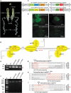
Figure 1.
Figure 1.
Genomic disruption of let-7 and let-7C by CRISPR/Cas9. (A) Morphology and divisions of the silk gland at D7 IL5. (B) The sgRNAs used for let-7 and let-7C knockout. The sequences in red are mature miRNAs, the sequences in green are the base-pairing regions of sgRNAs, and red boxes are protospacer-adjacent motifs (PAMs). (C) Schematic representation of the transgenic vectors for let-7 and let-7C knockout. Double-sgRNA expression cassettes were driven by the U6 promoter. EGFP was used as a selection marker. (D) Positive individuals selected at the G1 embryo and adult stages by screening the EGFP marker in the eye. (E) PCR products showing the chromosomal fragment deletion between sgRNA2 and sgRNA3 in Δlet-7-MSG. (F) PCR products of the chromosomal fragment deletion between sgRNA1 and sgRNA3 in Δlet-7C-MSG. (G–I) Sequence alignment of sgRNA-targeted genomic regions. The red arrow shows a fragment deletion.
Figure 2.
Figure 2.
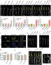
Knockout of let-7 and let-7C promoted PSG enlargement and improved silk yield. (A–D) Silk glands of Δlet-7-PSG and controls at D1 IL5, D3 IL5, D5 IL5 and D7 IL5. The red line shows the division between the MSG and the PSG. (E) Silk glands of Δlet-7C-PSG and controls at D7 IL5. (F,G) The length of the PSG was significantly increased in Δlet-7-PSG (F) and Δlet-7C-PSG (G) at D7 IL5. (H,I) The weight of the PSG was significantly increased in Δlet-7-PSG (H) and Δlet-7C-PSG (I) at D7 IL5. (J) The diameter of the PSG was increased in Δlet-7-PSG and Δlet-7C-PSG at D7 IL5. (K) Silk gland of Δlet-7-PSG post cocoon spinning or at pre-pupa stage. (L) Silk gland of Δlet-7C-PSG post cocoon spinning or at pre-pupa stage. (M) Cocoon weight of female and male was significantly increased in the Δlet-7-PSG and Δlet-7C-PSG. (N) Pupa weight of female and male was not altered in Δlet-7-PSG and Δlet-7C-PSG. (O) Phenotype difference between Δlet-7-PSG and Δlet-7C-PSG at D7 IL5. The error bars indicate the mean ± SEM, *P < 0.05, ***P < 0.001.
Figure 3.
Figure 3.
Transgenic overexpression of let-7 inhibited the development of PSG and the synthesis of fibroin protein. (A) Schematic diagram of the let-7 transgenic overexpression vector. (B) Schematic diagram of the miR-100+ miR-2795 transgenic overexpression vector. miRNA expression cassettes were driven by the FibH promoter. EGFP was used as a selection marker. (C) Silk gland of WT, let-7-OE-PSG and [miR-100+ miR-2795]-OE-PSG at D7 IL5. (D) Increased expression of let-7 in let-7-OE-PSG at D7 IL5. (E) Increased expression of miR-100 in [miR-100+ miR-2795]-OE-PSG at D7 IL5. (F) Increased expression of miR-2795 in [miR-100+ miR-2795]-OE-PSG at D7 IL5. (G) Length of the silk gland and its divisions decreased in the overexpression strain let-7-OE-PSG at D7 IL5. (H) The weight of cocoon with pupa increased in let-7-OE-PSG. (I) Cocoon shell weight decreased in let-7-OE-PSG. (J) Cocoon shell and pupa of let-7-OE-PSG and controls. (K) 20 mg cocoon shell of each strain was crushed and maintained in 8 M urea solution at 95 °C for one hour. (L) The weight of fibroin decreased in let-7-OE-PSG and [miR-100+ miR-2795]-OE-PSG. (M) The weight of female or male pupa increased in let-7-OE-PSG. (N) The length of female or male pupa increased in let-7-OE-PSG. The error bars indicate the mean ± SEM, *P < 0.01, **P < 0.05, ***P < 0.001.
Figure 4.
Figure 4.
Abnormal phenotypes caused by deletion of let-7 and let-7C in MSG. (A) The phenotypes of Δlet-7-MSG and Δlet-7C-MSG at D7 IL5. A, anterior region of MSG; M, middle region of MSG; P, posterior region of MSG. (B) The diameter of the P-MSG was increased in the Δ let-7-MSG and Δlet-7C-MSG mutants at D7 IL5. White: microscopy under common white light illumination; DAPI: fluorescent dye 4ʹ,6-diamidino-2-phenylindole and microscopy under fluorescence. (C) Silk glands of Δlet-7-MSG and Δlet-7C-MSG showing the residual sericin in the MSG lumen at prepupal stage. (D) The cocoon weight was significantly decreased in the Δlet-7-MSG and Δlet-7C-MSG mutants. The error bars indicate the mean ± SEM, **P < 0.01, ***P < 0.001.
Figure 5.
Figure 5.
Knockout of let-7 and let-7C promoted endoreduplication in silk gland cells. (A) The size of PSG cells increased dramatically in Δlet-7-PSG and Δlet-7C-PSG at D7 IL5. (B) DAPI staining showing DNA in the PSG of Δlet-7-PSG at D7 IL5. In (A) and (B), the red arrow shows the abnormal cell, and the dot line-enclosed area represents a single silk gland cell. (C) The DNA content increased in the P-MSG of Δlet-7-MSG and Δlet-7C-MSG at D7 IL5. (D) The DNA content increased in the M-MSG of Δlet-7-PSG and Δlet-7C-PSG at D7 IL5. (E) The ATP level increased in the M-MSG of Δlet-7-PSG and Δlet-7C-PSG at D3 IL5. The red arrow shows the malformed cell. (F) The volume of PSG cells decreased dramatically in the let-7-OE-PSG individuals at D7 IL5, but not in [miR-100+ miR-2795]-OE-PSG individuals at D7 IL5. (G) DAPI staining showing DNA in the PSG of let-7-OE-PSG at D7 IL5. In (F) and (G), the red and white arrows show the big and small cells, respectively. The error bars indicate the mean ± SEM, **P < 0.01, ***P < 0.001.
Figure 6.
Figure 6.
Identification of let-7 targets and transgenic overexpression of PC in MSG. (A) Schematic diagram of let-7 target screening. Venn diagram represents the overlap of potential targets predicted by three computational programs. Twenty-two of them are up-regulated by the let-7 deletion. (B) Two putative binding sites of let-7 within the 3ʹUTR of PC mRNA. Red: let-7 seed sequences; green, mutant target sites. (C) Dual-Luciferase Reporter Assay showing that let-7 directly targets the 3ʹ-UTR of PC by binding to two target sites in vitro. (D) Dual-Luciferase Reporter Assay showing that miR-100 and miR-2795 do not target the 3ʹ-UTR of PC in vitro. (E) let-7 deletion led to an increase of the PC transcript level in the MSG at D3 IL5. (F) Schematic recombinant vector for transgenic overexpression of PC gene in the silkworm MSG. (G) Screening of positive PC-OE-MSG strains at embryonic and adult stages. (a) Embryonic stage under the blue fluorescence. (b) Adult stage under the blue fluorescence. (H) The MSG enlarged at D7 IL5 after overexpression of PC. (I) PC expression levels increased in the MSG of PC-OE-MSG strain and miRNA mutants at D7 IL5. (J) Length of the MSG increased in the PC-OE-MSG strain at D7 IL5. (K) The DNA content increased in the MSG of PC-OE-MSG at D7 IL5. (L) Weight of larvae at D5 IL5. Error bars indicate the mean ± SEM, **P < 0.01, ***P < 0.001, ns represents not significant.
Figure 7.
Figure 7.
Regulatory network of let-7 is associated with the metabolism of glucose, fatty acids and amino acids in the silk gland. (A) Trehalose enters the silk gland cells and is converted into glucose. (B) let-7 knockout led to enhanced pentose phosphate pathway. (C) Knockout of let-7 promoted Embden-meyerhof pathway. (D) 3-phosphoglyceric acid was partly converted into amino acids as raw material for protein synthesis. (E) TCA cycle was promoted. (F) Gluconeogenesis pathway was weakened. (G) OAA and acetyl-CoA were replenished by CP cycle. (H) Synthesis metabolism of fatty acids was enhanced. (I) Fatty acid beta oxidation was enhanced. The red arrow shows the up-regulated gene and the blue arrow means the down-regulated gene. The abbreviations of Fig.7 are detailed in Table S3.

Similar articles

Cited by

References

    1. Xia Q, Guo Y, Zhang Z, et al. Complete resequencing of 40 genomes reveals domestication events and genes in silkworm (Bombyx). Science. 2009;326(5951):433–436. - PMC - PubMed
    1. Akai H. The structure and ultrastructure of the silk gland. Experientia. 1983;39:443–449.
    1. Xia Q, Li S, Feng Q. Advances in silkworm studies accelerated by the genome sequencing of Bombyx mori. Annu Rev Entomol. 2014;59:513–536. - PubMed
    1. Ma L, Xu H, Zhu J, et al. Ras1(CA) overexpression in the posterior silk gland improves silk yield. Cell Res. 2011;21(6):934–943. - PMC - PubMed
    1. Dong Z, Zhao P, Zhang Y, et al. Analysis of proteome dynamics inside the silk gland lumen of Bombyx mori. Sci Rep. 2016;6:21158. - PMC - PubMed

Publication types

MeSH terms