Case Reports
. 2020 Feb 4;20(1):46.
doi: 10.1186/s12886-020-1326-4.

Multimodal imaging in a case of a congenital retinal macrovessel associated with a retinal cavernous hemangioma: a case report

Affiliations
Case Reports

Multimodal imaging in a case of a congenital retinal macrovessel associated with a retinal cavernous hemangioma: a case report

Walid Zbiba et al. BMC Ophthalmol. .

Abstract

Background: To report the results of multimodal imaging in a case of a congenital retinal macrovessel associated with a retinal cavernous hemangioma.

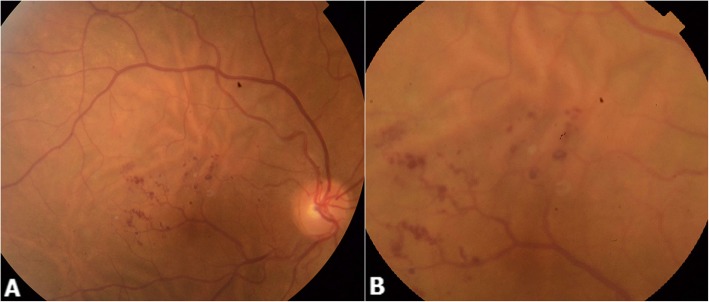
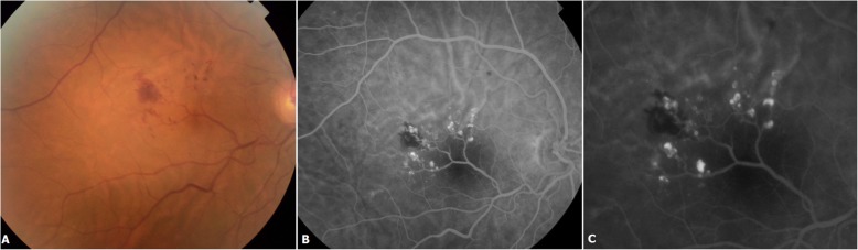
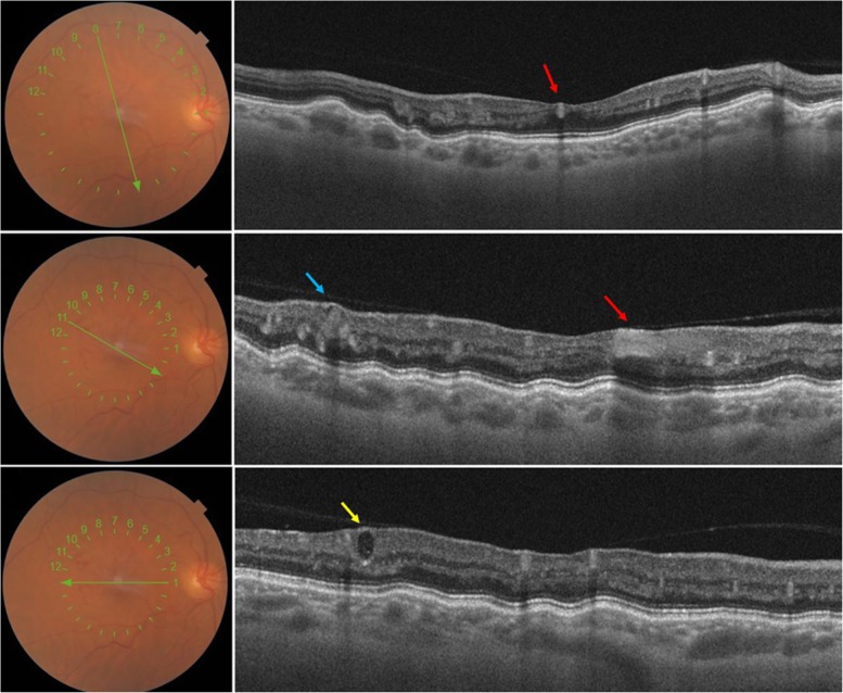
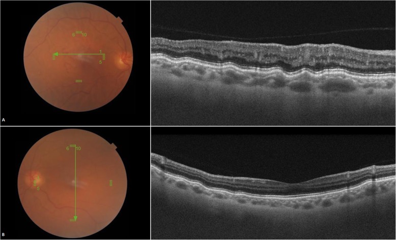
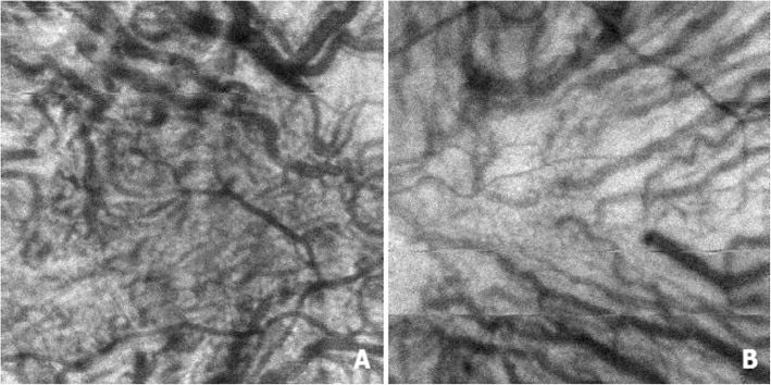
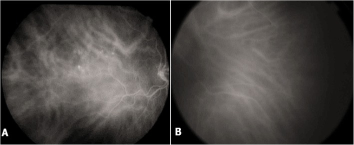
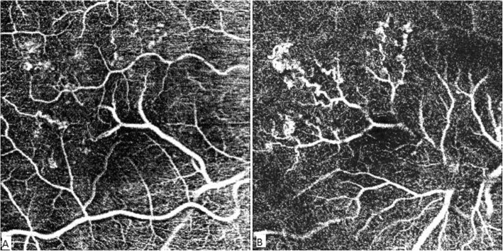
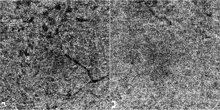
Case presentation: A 52-year-old female patient presented with progressive vision loss in the right eye. BCVA was 8/20 in the right eye and 18/20 in the left eye. Fundus examination of the right eye showed an aberrant retinal macrovessel arising from the inferior temporal major vein. It crossed the foveal area and overstepped to the superior retina. A "brunch of grapes" shaped retinal lesions arised from the macrovessel. Fluorescein angiography showed saccular lesions that filled slowly during the venous phase and became brightly hyperfluorescent saccular caps. SS-OCT of the right eye revealed a highly back-scattering hyper-reflective vessel across the fovea with shadow effect and adhesions between the vitreous and the aberrant macrovessel. It also revealed hypo reflective saccules with hyperreflective borders located in the inner retina corresponding to the cavernous retinal hemangioma. Retinal pigment epithelium undulations and vascular dilations at the level of Haller's layer were observed in both eyes. Indocyanine green angiography revealed chroidal vascular dilatations in both eyes in the late phase. OCT-A showed the aberrant vessel emerging from the inferior temporal vein and splitting the foveal avascular zone horizontally. RCH appeared as small heterogeneous saccular flow areas associated with focal capillary hypo perfusion areas. Asymmetry and distorsion of the foveal avascular zone were also noticed. A diagnosis of retinal macrovessel associated with a retinal cavernous hemangioma was made.

Conclusions: Congenital retinal macrovessels and retinal cavernous hemangioma are benign lesions. Their association is rare. Abnormal vascular development is likely to be responsible for their association. Swept source OCT and OCT angiography may be of a great contribution to better evaluate these retinal vascular disorders.

Keywords: Cavernous hemangioma; Chorioretinal folds; Congenital macrovessel; Optical coherence tomography angiography.


Conflict of interest statement

The authors declare that they have no competing interests.

Figures

Fig. 1
Fig. 1
Color fundus photography of the right eye (a and b) showing an aberrant retinal macrovessel arising from the inferior temporal major vein, crossing the foveal area, and oversteppping to the superior retina (a). A two optic disc diameter sized “brunch of grapes” shaped retinal lesions arise from the aberrant macrovessel (b). Dilated choroidal vessels with choroidal folds also are visible. (a and b)
Fig. 2
Fig. 2
Color fundus photography showing an intra-retinal hemorrhage located temporally to the fovea (a). Fluorescein angiography showing saccular lesions that fill slowly during the venous phase (b) and become brightly hyperfluorescent saccular caps with no leakage (c). The aberrant vessel’s filling is normal (b)
Fig. 3
Fig. 3
Optical coherence tomography (B scan) of the right eye showing highly back-scattering hyper-reflective vessel crossing the fovea (red arrow) with shadow effect (B) and adhesions between the posterior hyaloid and the aberrant macrovessel (B). Hypo reflective saccules with hyper reflective borders located in the inner retina corresponding to the cavernous retinal hemangioma (CRH) (blue arrow). Variable degrees of hypo and hyper reflectivities were observed inside these cystic lesions (Yellow arrow)
Fig. 4
Fig. 4
Optical coherence tomography (B scan) showing retinal pigment epithelium undulations in both eyes (a, b) more pronounced in the right eye (a), choroidal thickness is within normal limits, and there are vascular dilations at the level of Haller’s layer in both eyes more marked in the right eye
Fig. 5
Fig. 5
En face OCT of the right eye (a), left eye (b) showing bilateral choroidal vascular dilatations
Fig. 6
Fig. 6
Indocyanine green angiography of the right eye (a), left eye (b) showing bilateral choroidal vascular dilatations more marked in the right eye. Aneurysmal lesions are present in the right eye (a)
Fig. 7
Fig. 7
OCT-A of the superficial plexus (SCP) (a) showing the aberrant vessel emerging from the inferior temporal vein and branching at the level of the fovea. It splits the foveal avascular zone (FAZ) horizontally and causes an asymmetry and distortion of the FAZ (a). The terminal branches of the aberrant vessels are observed at the level of the deep capillary plexus (DCP) (b). Retinal cavernous hemangiomas (RCH) appear as small heterogeneous saccular flow areas identified on the deep plexus along the branches of the macrovessel (b). Hypo perfusion areas surrounding the vascular anomalies are visible in both the SCP and DCP (a and b)
Fig. 8
Fig. 8
Right eye OCT-A image of choriocapillaris illustrates multifocal hypointense areas that precisely colocalize with the aberent vessel and the cavernous hemangiomas (a). Flow in the Choriocapillaris was normal in the left eye (b)

References

    1. Strampe MR, Wirostko WJ, Carroll J. A case of congenital retinal macrovessel in an otherwise normal eye. Am J Ophthalmol Case Rep. 2017;8:18–21. doi: 10.1016/j.ajoc.2017.09.001. - DOI - PMC - PubMed
    1. Goel N, Kumar V, Seth A, Ghosh B. Branch retinal artery occlusion associated with congenital retinal macrovessel. Oman J Ophthalmol. 2014;7(2):96–97. doi: 10.4103/0974-620X.137172. - DOI - PMC - PubMed
    1. Bhatia HK. Congenital retinal macrovessel with normal visual acuity: a case report. Int J Ophthalmolo Clin Res. 2015;2(2):017.
    1. Sanfilippo CJ, Sarraf D. Congenital macrovessel associated with cystoid macular edema and an ipsilateral intracranial venous malformation. Retin Cases Brief Rep. 2015;9(4):357–359. doi: 10.1097/ICB.0000000000000236. - DOI - PMC - PubMed
    1. Thanos A, Randhawa S, Drenser KA. Macular retinal cavernous hemangioma associated with congenital retinal macrovessel. JAMA Ophthalmol. 2016;134(9):e161683. doi: 10.1001/jamaophthalmol.2016.1683. - DOI - PubMed

Publication types

LinkOut - more resources