. 2020 Feb 4;8(1):13.
doi: 10.1186/s40478-020-0884-2.

Pre-clinical characterisation of E2814, a high-affinity antibody targeting the microtubule-binding repeat domain of tau for passive immunotherapy in Alzheimer's disease

Affiliations

Pre-clinical characterisation of E2814, a high-affinity antibody targeting the microtubule-binding repeat domain of tau for passive immunotherapy in Alzheimer's disease

Malcolm Roberts et al. Acta Neuropathol Commun. .

Abstract

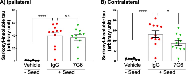
Tau deposition in the brain is a pathological hallmark of many neurodegenerative disorders, including Alzheimer's disease (AD). During the course of these tauopathies, tau spreads throughout the brain via synaptically-connected pathways. Such propagation of pathology is thought to be mediated by tau species ("seeds") containing the microtubule binding region (MTBR) composed of either three repeat (3R) or four repeat (4R) isoforms. The tau MTBR also forms the core of the neuropathological filaments identified in AD brain and other tauopathies. Multiple approaches are being taken to limit tau pathology, including immunotherapy with anti-tau antibodies. Given its key structural role within fibrils, specifically targetting the MTBR with a therapeutic antibody to inhibit tau seeding and aggregation may be a promising strategy to provide disease-modifying treatment for AD and other tauopathies. Therefore, a monoclonal antibody generating campaign was initiated with focus on the MTBR. Herein we describe the pre-clinical generation and characterisation of E2814, a humanised, high affinity, IgG1 antibody recognising the tau MTBR. E2814 and its murine precursor, 7G6, as revealed by epitope mapping, are antibodies bi-epitopic for 4R and mono-epitopic for 3R tau isoforms because they bind to sequence motif HVPGG. Functionally, both antibodies inhibited tau aggregation in vitro. They also immunodepleted a variety of MTBR-containing tau protein species. In an in vivo model of tau seeding and transmission, attenuation of deposition of sarkosyl-insoluble tau in brain could also be observed in response to antibody treatment. In AD brain, E2814 bound different types of tau filaments as shown by immunogold labelling and recognised pathological tau structures by immunohistochemical staining. Tau fragments containing HVPGG epitopes were also found to be elevated in AD brain compared to PSP or control. Taken together, the data reported here have led to E2814 being proposed for clinical development.

Keywords: Alzheimer; Tau; immunotherapy; neurodegeneration; tauopathy.


Conflict of interest statement

All work performed to generate data reported in this manuscript was funded by Eisai, a pharmaceutical company listed on the Tokyo Stock Exchange (TYO:4523). MR, YI, KM, ST, MD, JG, HO, ZZ, SA, NT, MO, MA, HA, KLA, JS, EA, KH and JMS were full-time employees of Eisai for the period in which data reported in this study were generated. IS and ES are UCL employees funded by Eisai through the Eisai:UCL Therapeutic Innovation Group (TIG). The TIG is a collaborative partnership between Eisai and UCL to discover and advance medical therapies for neurodegenerative disorders. Successful medical therapies from the collaboration could potentially be of commercial benefit to both Eisai and UCL. RdS and KS are UCL employees and also funded by the Reta Lila Weston Trust for Medical Research.

Figures

Fig. 1
Fig. 1
Peptide selection for antibody generation. A schematic representation of all six tau isoforms expressed in human adult brain is shown. The two hexapeptide sequences necessary to initiate tau aggregation, PHF6* and PHF6, are also indicated. To generate potential therapeutic antibodies, two peptide immunogens contained within the microtubule-binding region (MTBR) of 4R tau isoforms were selected. Peptide 1 (273–291) contained two amino acids within the R1 region with the remainder in R2 encompassing the PHF6* motif. Peptide 2 (296–314) contained ten amino acids within R2 and nine in R3, also including the PHF6 motif as well as the P301 residue (bold and underlined), often introduced as mutated in pre-clinical research models. Amino-acid numbering according to largest (2N4R) isoform (NP_005901)
Fig. 2
Fig. 2
Fine epitope mapping of 7G6 and E2814 anti-tau antibodies. Overlapping peptides covering the full length wild type human 2N4R tau sequence were synthesised and printed onto glass chips. Each chip was then probed with either a) 7G6 (mouse) or b) E2814 (human) antibodies at 1 μg/mL. Bound antibodies were detected by the addition of fluorescently-labelled secondary antibodies and images captured using a LI-COR Odyssey machine. Fluorescent spots corresponding to antibody-bound peptides were quantified and intensity plots spanning the full tau sequence were generated for both murine 7G6 (a) and human E2814 (b) antibodies. Both antibodies bound peptides containing an HVPGG sequence as indicated in each panel
Fig. 3
Fig. 3
Immunohistochemical staining of tauopathy sections with E2814. a-c: Frontal cortex from AD brain sections stained with 0.5 μg/mL E2814 showing robust labelling of neurofibrillary tangles (arrows). Nuclei stained in blue (quad arrow); d, e: Control staining of AD brain sections with human IgG also at 0.5 μg/mL. f-h,j,k: Frontal cortex from a PSP patient stained with E2814 (0.2 μg/mL) showing widespread staining of neurofibrillary tangles (arrows) and glial inclusions, including tufted astrocytes (G,H; arrowheads) and oligodendroglial coiled body (k; arrow). l: Control staining of a PSP section with human IgG (0.33 μg/mL). Non-specific brown colouration is due to lipofuscin. Nuclei are stained in blue (quad arrows); m,n: Hippocampus from a PiD patient stained with E2814 (0.2 μg/mL) showing strong staining of Pick bodies (arrows). p: Control staining of PiD brain section with human IgG (0.33 μg/mL). Nuclei are stained in blue (quad arrows). Scale bars = 50 μm
Fig. 4
Fig. 4
Immunogold labelling of tau filaments from human AD brain. Representative electron microscope images of tau fibrils isolated from the sarkosyl-insoluble fraction of AD patient frontal cortex. E2814 or IgG1 isotype control were used at 10 μg/mL. Tau 5 antibody was used at 0.4 μg/mL. Bound antibody was detected following addition of an anti-human 12 nm gold conjugated antibody at 1:25 dilution or an anti-mouse 6 nm gold conjugated antibody at 1:25 dilution (for Tau 5). a, b: E2814 could bind the entire length of many tau fibrils. In some paired helical filaments (PHFs) E2814 binding was limited to the ends (arrows) of the fibrils (c) or was completely absent (d). f: E2814 (arrows) and the commercially available Tau 5 antibody (arrowheads) co-stain tau fibrils, providing proof of E2814 specificity to tau fibrils. g: Tau 5 binds to the entire length of tau fibrils. h: E2814 specifically binds to smaller structures on the EM grids that may represent tau fibril fragments or tau oligomers (arrows). The IgG1 control antibody did not bind to filbrils (e), or smaller fragments (f). j,k). Pre-treatment of AD fibrils with 0.4 mg/ml pronase removed the fuzzy coat of PHFs and SFs, leaving the structured core intact with E2814 staining retained only at some fibril ends (Scale bars = 200 nm)
Fig. 5
Fig. 5
Inhibition of tau aggregation in vitro. Recombinant wild-type (a) or P301S mutant (b) tau at 12 μM was induced to aggregate in vitro with addition of heparin in the absence or presence of either 7G6, E2814 or control IgG1 antibodies at a concentration of 8.3 μM. Over a time course of 6 days, samples of the reaction mixture were removed and incubated with Thioflavin S (ThS) and fluorescence was measured to detect aggregated tau. Data shown represent six independent experiments for each protein. A two-way ANOVA statistical analysis was performed followed by a Dunnett’s test. **** p ≤ 0.0001 7G6 or E2814 versus IgG, *** p ≤ 0.001 7G6 vs IgG for wild type protein only. Values represent mean ± SEM
Fig. 6
Fig. 6
Immunodepletion of tau seeds with E2814 reduces intracellular tau deposition. E2814 antibody or human IgG1 control at specified concentrations were used to immunodeplete K18 fibrils (a,b) or full length P301S tau monomer (c,d) seeds. Treated samples were added to HEK293 cells overexpressing P301S mutant 0N4R tau. Intracellular tau deposition was measured by addition of Thioflavin S (ThS) and cells were counterstained with DAPI to visualise the nuclei. The percentage of ThS/DAPI relative to IgG1 control is plotted (IgG1 = 100% seeding effect). Values represent the mean ± SEM from four (a) and three (c) independent experiments. Data were analysed by one way ANOVA followed by Dunnett’s test. *P < 0.05, **P < 0.005, **** < 0.0001. Representative images of ThS and DAPI staining for K18 fibril (B, 3 μg antibody) and P301S monomer (d, 10 μg antibody) immunodepleted seeds are shown. Scale bars = 200 μm
Fig. 7
Fig. 7
7G6 efficacy in an in vivo model of tau seeding and transmission. Full-length P301S tau seeds were injected into the left hippocampus of P301S transgenic mice pretreated with either IgG control (n = 11) or 7G6 antibody (n = 11) at a dose of 40 mg/kg i.p. Animals then received the same dose of antibody or vehicle once per week for a further 3 weeks until sacrifice. Vehicle-treated animals receiving no seed (n = 6) were included as an additional control group. Both hippocampi from each brain were extracted and separately treated with sarkosyl. Tau levels were then quantified in the sarkosyl-insoluble fraction from each sample by western blot and plotted for either the ipsilateral (a) or contralateral (b) hippocampi for each animal. Data are expressed as mean ± SEM and further analysed using a one-way ANOVA followed by Fisher’s LSD test. **** p ≤ 0.0001 IgG versus no seed control, * p ≤ 0.05 7G6 versus IgG for contralateral hippocampus, n.s. not significant
Fig. 8
Fig. 8
LC/MS analysis of tau peptide abundance in the insoluble fraction from human tauopathy brain. Insoluble fractions from AD (n = 3), PSP (n = 4) and control (n = 2) brain samples were prepared and digested with Lys-c and trypsin. Tryptic peptides were identified and quantified by LC/MS. The standardised abundance for each tau peptide was calculated (see Materials and Methods) and compared between AD, PSP and control brain samples. Peptides showing significant change between PSP and AD are plotted. Error bars for AD and PSP represent standard deviation
Fig. 9
Fig. 9
Size exclusion chromatography of tauopathy brain samples. Human brain homogenates from AD (upper panels) or PSP (lower panels) patients were loaded onto a Superdex 200 Increase GL size exclusion column in PBS and 1 mL fractions were collected. Samples from each fraction were resolved by SDS-PAGE and tau proteins were detected by western blotting using antibodies raised against two different parts of the full length protein: HT7 (0.2 μg/mL) or 7G6 (0.5 μg/mL) as indicated in the left or right panels, respectively. Images shown are representative from four different AD and six PSP brains
Fig. 10
Fig. 10
AD tau protofibril structure (modified from [26]): Schematic representation of protein backbone of the R3 + R4 protofibril unit of AD tau paired-helical filaments and straight filaments, with selected amino acid side chains. The numbering of amino acid positions is based on the 2N4R-tau isoform (NP_005901). Antiparallel β-strands are indicated by thick arrows. In AD, the protofibril adopts a compact C-shaped structure. The E2814 HVPGG binding motif (362-366; yellow) forms the tight bend between β7 and β8. Red arrowheads indicate predicted strong contact points to heparin [53] resulting in heparin-mediated aggregation by compaction and stabilization of the AD protofibril. Green stars indicate sequences that interact with azure A and azure B, monodemethylated derivatives of methylene blue (MB), which have anti-aggregation effects on tau by preventing fibril formation and retaining tau in monomeric form [2]

References

    1. Ahmed Z, Cooper J, Murray TK, Garn K, McNaughton E, Clarke H, Parhizkar S, Ward MA, Cavallini A, Jackson S, et al. A novel in vivo model of tau propagation with rapid and progressive neurofibrillary tangle pathology: the pattern of spread is determined by connectivity, not proximity. Acta Neuropathol. 2014;127:667–683. doi: 10.1007/s00401-014-1254-6. - DOI - PMC - PubMed
    1. Akoury E, Pickhardt M, Gajda M, Biernat J, Mandelkow E, Zweckstetter M. Mechanistic basis of phenothiazine-driven inhibition of Tau aggregation. Angew Chem Int Ed Engl. 2013;52:3511–3515. doi: 10.1002/anie.201208290. - DOI - PubMed
    1. Allen B, Ingram E, Takao M, Smith MJ, Jakes R, Virdee K, Yoshida H, Holzer M, Craxton M, Emson PC, et al. Abundant tau filaments and nonapoptotic neurodegeneration in transgenic mice expressing human P301S tau protein. J Neurosci. 2002;22:9340–9351. doi: 10.1523/JNEUROSCI.22-21-09340.2002. - DOI - PMC - PubMed
    1. Arai T, Ikeda K, Akiyama H, Nonaka T, Hasegawa M, Ishiguro K, Iritani S, Tsuchiya K, Iseki E, Yagishita S, et al. Identification of amino-terminally cleaved tau fragments that distinguish progressive supranuclear palsy from corticobasal degeneration. Ann Neurol. 2004;55:72–79. doi: 10.1002/ana.10793. - DOI - PubMed
    1. Asuni AA, Boutajangout A, Quartermain D, Sigurdsson EM. Immunotherapy targeting pathological tau conformers in a tangle mouse model reduces brain pathology with associated functional improvements. J Neurosci. 2007;27:9115–9129. doi: 10.1523/JNEUROSCI.2361-07.2007. - DOI - PMC - PubMed

Publication types

MeSH terms