. 2020 Feb 4;11(1):e02699-19.
doi: 10.1128/mBio.02699-19.

The Toxoplasma gondii Cyst Wall Interactome

Affiliations

The Toxoplasma gondii Cyst Wall Interactome

Vincent Tu et al. mBio. .

Abstract

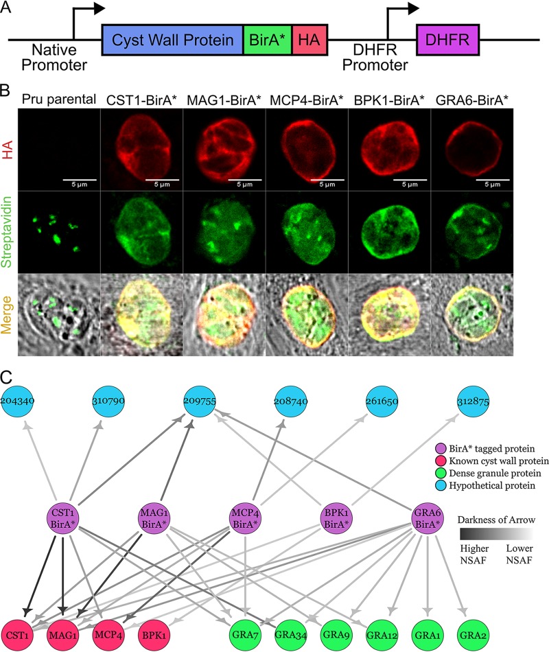
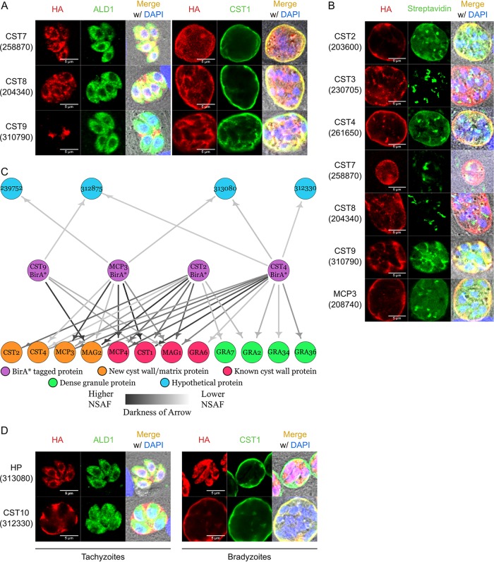
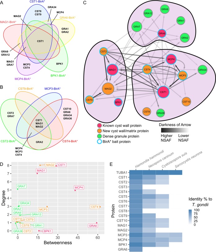
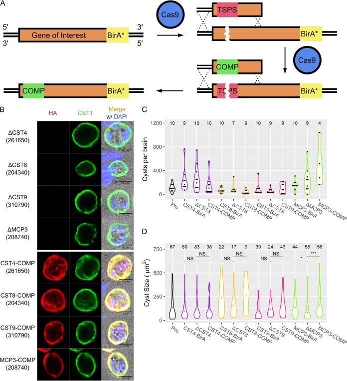
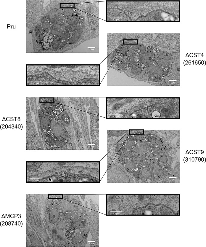
A characteristic of the latent cyst stage of Toxoplasma gondii is a thick cyst wall that forms underneath the membrane of the bradyzoite vacuole. Previously, our laboratory group published a proteomic analysis of purified in vitro cyst wall fragments that identified an inventory of cyst wall components. To further refine our understanding of the composition of the cyst wall, several cyst wall proteins were tagged with a promiscuous biotin ligase (BirA*), and their interacting partners were screened by streptavidin affinity purification. Within the cyst wall pulldowns, previously described cyst wall proteins, dense granule proteins, and uncharacterized hypothetical proteins were identified. Several of the newly identified hypothetical proteins were validated to be novel components of the cyst wall and tagged with BirA* to expand the model of the cyst wall interactome. Community detection of the cyst wall interactome model revealed three distinct clusters: a dense granule, a cyst matrix, and a cyst wall cluster. Characterization of several of the identified cyst wall proteins using genetic strategies revealed that MCP3 affects in vivo cyst sizes. This study provides a model of the potential protein interactions within the cyst wall and the groundwork to understand cyst wall formation.IMPORTANCE A model of the cyst wall interactome was constructed using proteins identified through BioID. The proteins within this cyst wall interactome model encompass several proteins identified in a prior characterization of the cyst wall proteome. This model provides a more comprehensive understanding of the composition of the cyst wall and may lead to insights on how the cyst wall is formed.

Keywords: BirA; Toxoplasma gondii; cyst wall; protein interactions; proteomics.


Figures

FIG 1
FIG 1
Initial cyst wall pulldowns with BioID reveal hypothetical proteins. (A) Schematic diagram of the exogenous expression construct containing the promoter and coding sequence of the cyst wall gene of interest fused C terminally with BirA* and 3×HA followed by the DHFR selectable marker. (B) Immunofluorescence micrographs of Pru and BirA*-tagged parasites (CST1, MAG1, MCP4, BPK1, and GRA6) stained with anti-HA antibody and Alexa Fluor 488-conjugated streptavidin under pH 8 conditions supplemented with 150 μM biotin. Endogenously biotinylated proteins within the apicoplast are observed within these parasite strains. (C) Network graph showing potential parasite-specific interacting proteins of each BirA*-tagged cyst wall protein (CST1, MAG1, MCP4, BPK1, and GRA6) identified during the bradyzoite stage. BirA* cyst wall bait proteins, known cyst wall proteins, dense granule proteins, and hypothetical proteins are represented by purple, red, green, and blue nodes, respectively. Darker arrows correspond to higher normalized spectral abundance factor (NSAF) values of prey proteins identified in each pulldown.
FIG 2
FIG 2
Iterative BioID pulldowns of cyst wall proteins identify a novel cyst wall protein. (A) Immunofluorescence images of tagged hypothetical proteins stained with anti-HA and anti-ALD1 (cytosolic marker) or anti-CST1 (cyst wall marker) antibodies. Tachyzoites: HA in red and ALD1 in green; bradyzoites: HA in red and CST1 in green. (B) Immunofluorescence micrographs of endogenously BirA*-tagged parasites (CST2, CST3, CST4, CST7, CST8, CST9, and MCP3) stained with anti-HA antibody and Alexa Fluor 488-conjugated streptavidin under pH 8 conditions supplemented with 150 μM biotin. (C) Network graph showing potential parasite-specific interacting proteins of each BirA*-tagged cyst wall protein (CST2, CST4, CST9, and MCP3). BirA* cyst wall bait proteins, known cyst wall proteins, novel cyst wall proteins, dense granule proteins, and hypothetical proteins are represented by purple, red, orange, green, and blue nodes, respectively. Darker arrows correspond to higher normalized spectral abundance factor (NSAF) values of prey proteins identified in each pulldown. (D) Immunofluorescence images of HA-tagged hypothetical proteins (HP) identified from the group 2 BirA* pulldowns stained with anti-HA and anti-ALD1 (cytosolic marker) or anti-CST1 (cyst wall marker) antibodies. Tachyzoites: HA in red and ALD1 in green; bradyzoites: HA in red and CST1 in green.
FIG 3
FIG 3
Cyst wall interactome model analysis reveals distinct clusters. Venn diagrams showing the proteins that are common between the group 1 (A) or group 2 (B) cyst wall BirA* pulldowns. Each BirA* pulldown is represented by a different colored circle. (C) Network of the proteins identified in the group 1 and group 2 BirA* pulldowns. BirA* bait protein nodes are represented by a blue outline. Known cyst wall proteins, dense granule proteins, and novel cyst wall/matrix proteins are represented by red, green, and orange nodes, respectively. Darkness of arrows represents the normalized spectral abundance factor (NSAF) of the prey proteins identified in each pulldown. Size of each node represents the in-degrees or the amount of times a protein was identified within each pulldown. Protein clustering was performed using the Louvain method of community detection. (D) Graph of each node’s betweenness, which signifies the extent to which nodes stand between each other, versus their in-degrees, the frequency of each node appearing in a pulldown. (E) Heat map showing the percentage of identity between alpha tubulin and each T. gondii cyst wall and cyst matrix protein to their orthologs in other cyst-forming coccidians.
FIG 4
FIG 4
Characterization of cyst wall proteins reveals MCP3 affects cyst size. (A) Schematic showing the endogenous editing of genomic loci using a targetable stop PAM sequence (TSPS) to introduce a Cas9-targetable sequence of premature stop codons. Complementation of the edited loci was performed by targeting the premature stop codons with a TSPS-specific sgRNA and replacing the TSPS with synonymous mutations of the coding region. The red region indicates the TSPS sequence, while the green region indicates synonymous mutations. (B) IFA showing CST4, CST8, CST9, and MCP3 knockout (top) and complement (bottom) bradyzoites stained with anti-HA (red) and anti-CST1 (green) antibodies. (C) Brain cyst counts of mice infected with Pru, CST4, CST8, CST9, and MCP3 knockout and complement parasite strains at 30 days postinfection. Numbers of brains counted are listed above each violin plot. No significant differences in cyst numbers between BirA*, knockout, and complement parasites in each strain group were observed (P > 0.05). (D) Size analysis of in vivo cysts obtained from Pru, CST4, CST8, CST9, and MCP3 knockout and complement parasite strains. Numbers of cysts imaged and analyzed are listed above each violin plot. NS, not significant; *, P < 0.05; ***, P < 0.001.
FIG 5
FIG 5
Novel cyst wall proteins do not affect morphology of cyst ultrastructure. Transmission electron micrographs of in vitro cysts from the Pru, ΔCST4, ΔCST8, ΔCST9, and ΔMCP3 parasite strains.

References

    1. Porter SB, Sande MA. 1992. Toxoplasmosis of the central nervous system in the acquired immunodeficiency syndrome. N Engl J Med 327:1643–1648. doi:10.1056/NEJM199212033272306. - DOI - PubMed
    1. Antczak M, Dzitko K, Długońska H. 2016. Human toxoplasmosis-searching for novel chemotherapeutics. Biomed Pharmacother 82:677–684. doi:10.1016/j.biopha.2016.05.041. - DOI - PubMed
    1. Lemgruber L, Lupetti P, Martins-Duarte ES, De Souza W, Vommaro RC. 2011. The organization of the wall filaments and characterization of the matrix structures of Toxoplasma gondii cyst form. Cell Microbiol 13:1920–1932. doi:10.1111/j.1462-5822.2011.01681.x. - DOI - PubMed
    1. Parmley SF, Yang S, Harth G, Sibley LD, Sucharczuk A, Remington JS. 1994. Molecular characterization of a 65-kilodalton Toxoplasma gondii antigen expressed abundantly in the matrix of tissue cysts. Mol Biochem Parasitol 66:283–296. doi:10.1016/0166-6851(94)90155-4. - DOI - PubMed
    1. Buchholz KR, Fritz HM, Chen X, Durbin-Johnson B, Rocke DM, Ferguson DJ, Conrad PA, Boothroyd JC. 2011. Identification of tissue cyst wall components by transcriptome analysis of in vivo and in vitro Toxoplasma gondii bradyzoites. Eukaryot Cell 10:1637–1647. doi:10.1128/EC.05182-11. - DOI - PMC - PubMed

Publication types

LinkOut - more resources