. 2020 Mar 13;295(11):3518-3531.
doi: 10.1074/jbc.RA119.011332. Epub 2020 Feb 4.

A CLOCK-binding small molecule disrupts the interaction between CLOCK and BMAL1 and enhances circadian rhythm amplitude

Affiliations

A CLOCK-binding small molecule disrupts the interaction between CLOCK and BMAL1 and enhances circadian rhythm amplitude

Yagmur Umay Doruk et al. J Biol Chem. .

Abstract

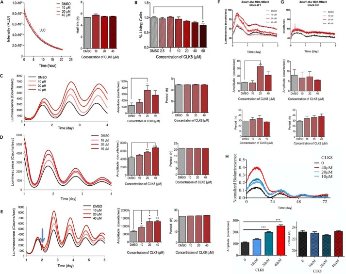
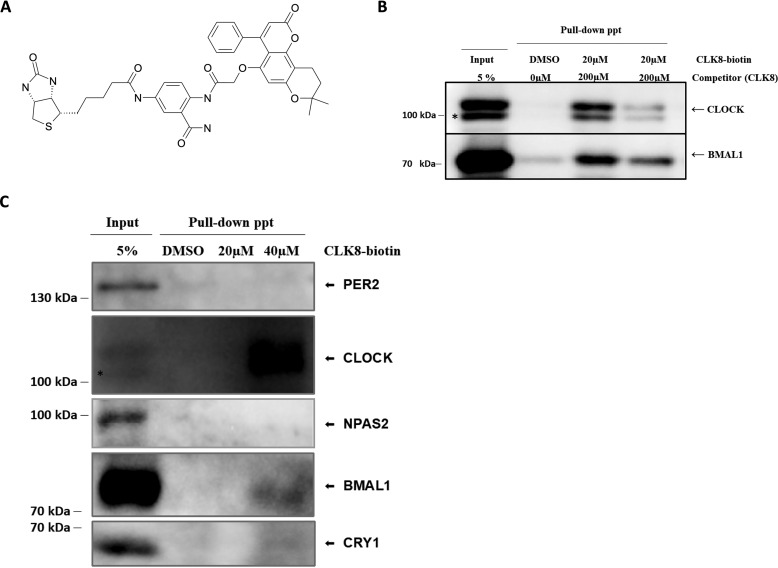
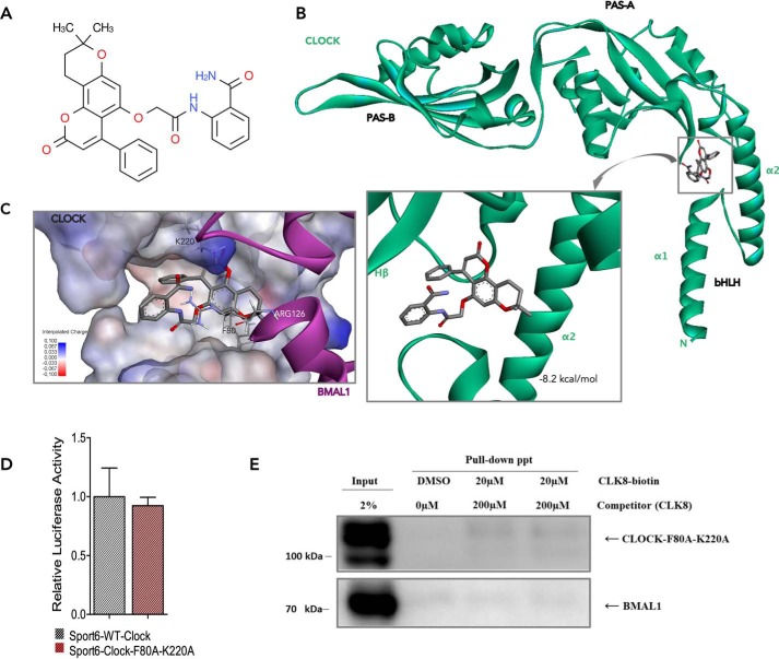
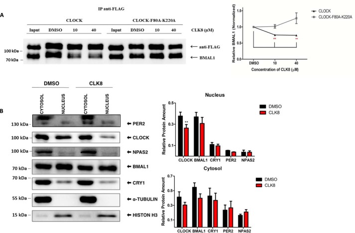
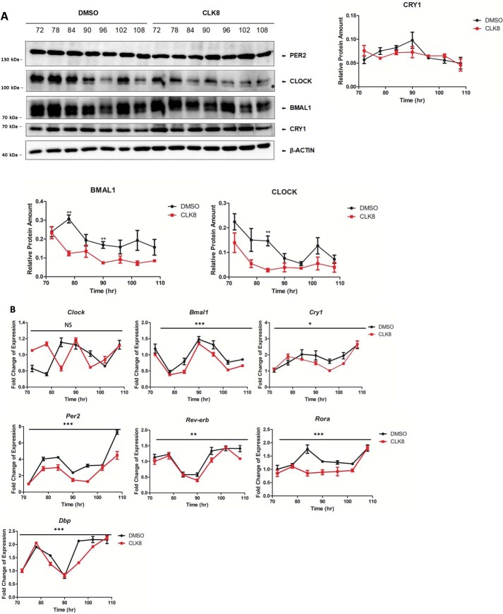
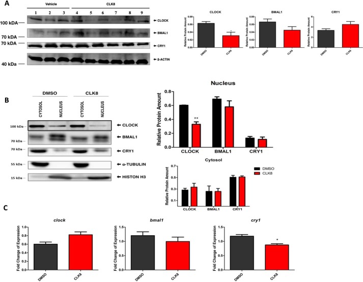
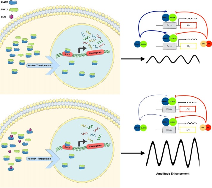
Proper function of many physiological processes requires a robust circadian clock. Disruptions of the circadian clock can result in metabolic diseases, mood disorders, and accelerated aging. Therefore, identifying small molecules that specifically modulate regulatory core clock proteins may potentially enable better management of these disorders. In this study, we applied a structure-based molecular-docking approach to find small molecules that specifically bind to the core circadian regulator, the transcription factor circadian locomotor output cycles kaput (CLOCK). We identified 100 candidate molecules by virtual screening of ∼2 million small molecules for those predicted to bind closely to the interface in CLOCK that interacts with its transcriptional co-regulator, Brain and muscle Arnt-like protein-1 (BMAL1). Using a mammalian two-hybrid system, real-time monitoring of circadian rhythm in U2OS cells, and various biochemical assays, we tested these compounds experimentally and found one, named CLK8, that specifically bound to and interfered with CLOCK activity. We show that CLK8 disrupts the interaction between CLOCK and BMAL1 and interferes with nuclear translocation of CLOCK both in vivo and in vitro Results from further experiments indicated that CLK8 enhances the amplitude of the cellular circadian rhythm by stabilizing the negative arm of the transcription/translation feedback loop without affecting period length. Our results reveal CLK8 as a tool for further studies of CLOCK's role in circadian rhythm amplitude regulation and as a potential candidate for therapeutic development to manage disorders associated with dampened circadian rhythms.

Keywords: brain and muscle Arnt-like protein-1 (BMAL1); circadian clock; circadian locomotor output cycles kaput (CLOCK); circadian regulation; circadian rhythm amplitude; drug design; drug development; gene expression; gene regulation; protein expression; transcription; transcription coregulator.


Conflict of interest statement

The authors declare that they have no conflicts of interest with the contents of this article

Figures

Figure 1.
Figure 1.
Initial screening for identification of CLK8. A, HEK293T cells were transfected with pcDNA-Luc and the effect of 10, 20, and 40 μm CLK8 on the luciferase (LUC) half-life was measured. The Luc signal was monitored after CLK8 treatment until it reached a plateau. Data are mean ± S.E.; n = 3 independent experiments. B, dose-dependent cytotoxicity of CLK8 in the U2OS cells. All the measurements were normalized to 0.5% DMSO control. Data are mean ± S.E.; n = 3 independent experiments. C and D, continuous monitoring of the luminescence rhythms of Bmal1-dLuc U2OS and Bmal1-dLuc NIH 3T3-transfected cells to determine the dose-dependent effect of CLK8 on circadian rhythm. Amplitude and period parameters are shown in the right panel. E, Bmal1-dLuc U2OS cells were synchronized at time 0. Two days later (blue arrow), cells were treated with 10, 20, or 40 μm CLK8. Cells treated with 0.5% DMSO were used as the control. Luminescence profiles are the means of three independent experiments. Amplitude and period parameters (shown in the right panel) were obtained by fitting first-order polynomial baseline-subtracted data with sin (damped). Day 1 was not included in the analysis. Data are mean ± S.E.; n = 3 independent experiments. *, p value <0.05; **, p value <0.01; ***, p value <0.001. F, WT MDA MB231 cells and G, CLOCK knock-out MDA MB231 cells were transduced with Bmal1-dluc lentiviral particles. At 72 h post-transduction, the luminescence rhythms of the cells were monitored continuously to determine the dose-dependent effect of CLK8 on circadian rhythm. Amplitude and period parameters (shown in the bottom panel) were obtained by fitting first-order polynomial baseline-subtracted data with sin (damped). Luminescence profiles are means of three independent experiments. Data are mean ± S.E.; n = 3 independent experiments. *, p value <0.05. H, primary mouse skin fibroblast cells were transduced with Bmal1-dluc lentiviral particles. Day 1 was not included in the analysis. Data are mean ± S.E.; n = 4 independent experiments. ***, p value <0.001.
Figure 2.
Figure 2.
Specific binding of CLK8 to CLOCK. A, chemical structure of biotinylated CLK8 (bait). Pulldown assay using whole cell lysates of (B) HEK293T cells overexpressing Clock and Bmal1 from pSport6 plasmids and (C) U2OS cells expressing endogenous levels of CLOCK (and BMAL1). Free CLK8 was used as the competitor. Data are representative of three independent experiments. Asterisk indicates nonspecific band.
Figure 3.
Figure 3.
Docking of CLK8 into the CLOCK-binding site. A, chemical structure of CLK8. B, best binding mode of CLK8 to CLOCK (green) with a predicted binding energy of −8.2 kcal/mol. CLK8 can enter the hollow between the α2 helix of the bHLH domain and the Hβ strand of the PAS-A domain of CLOCK. C, superposition of CLK8 and Arg-126 of BMAL1 (magenta). CLK8 and Arg-126 of BMAL1 share the same binding region in the CLOCK structure, where Phe-80 plays an essential role. The positively charged Lys-220 in the PAS-A domain of CLOCK also contributes to a π-cation interaction with CLK8. D, comparison of WT CLOCK and mutant CLOCK-F80A,K220A. The transcriptional functions of WT and mutant CLOCK were compared by a Per1-Luc assay in HEK293T cells. Data are mean ± S.E.; n = 3 independent experiments. E, pulldown assay using whole cell lysate of HEK293T cells overexpressing CLOCK-F80A,K220A and BMAL1.
Figure 4.
Figure 4.
Reduction of CLOCK and BMAL1 interaction and nuclear localization of CLOCK by CLK8. A, effect of CLK8 on the interaction of CLOCK and BMAL1 were evaluated by co-immunoprecipitation. Anti-FLAG affinity gel was used to precipitate FLAG-tagged CLOCK together or FLAG-tagged CLOCK-F80A,K220A mutant with BMAL1. Western blot analysis using anti-BMAL1 and anti-FLAG antibodies was performed to compare the association of CLOCK and the CLOCK-F80A,K220A mutant with BMAL1 in samples with different concentrations of CLK8. Quantification of the Western blotting is shown in the right panel. The vertical axis, which indicates the amount of BMAL1 (normalized by CLOCK) in each sample, was calculated relative to the DMSO control. B, unsynchronized U2OS cells were treated with 20 μm CLK8. Control cells were treated with 0.5% DMSO. Two days later, cells were fractionated and examined by Western blot analysis, where CLOCK, NPAS2, PER2, BMAL1, and CRY1 were normalized by α-tubulin or histone-H3. Quantifications are shown on the right. Data are mean ± S.E.; n = 5 independent experiments. The Western blotting data are representative of five independent experiments. *, p value <0.05; **, p value <0.01.
Figure 5.
Figure 5.
CLK8 altered CLOCK protein levels in a time-dependent manner. Confluent U2OS cells were synchronized by 2-h treatment with dexamethasone (0.1 μm) and the medium was replaced with fresh medium containing CLK8 or DMSO. Cells were harvested at the indicated time points. Asterisk indicates nonspecific band. A, time-dependent analysis of core clock proteins by Western blotting. Data are mean ± S.E.; n = 3. B, qPCR analysis of the core clock genes in a time-dependent manner. Data are the mean ± S.E.; n = 3 independent experiments. ***, p < 0.001; **, p < 0.005; *, p < 0.05 versus the DMSO control by two-way ANOVA.
Figure 6.
Figure 6.
Effect of CLK8 in mice liver. A, the mice were treated with CLK8 or vehicle and then euthanized. Liver protein lysates were prepared and probed with antibodies for different core clock proteins. B, mice livers (n = 3 for vehicle-treated animals; n = 5 for CLK8-treated animals) were fractionated and the samples were subjected to Western blot analysis. Quantifications are shown in the right panel. Data are mean ± S.E.; n = 3 independent experiments for the control samples; n = 5 independent experiments for CLK8-treated samples. **, p value <0.01 by one-way ANOVA. C, transcriptional levels of Clock, Cry1, and Bmal1 were measured by qPCR. Data are mean ± S.E.; n = 3 independent for the control samples; n = 5 for CLK8-treated samples. *, p value <0.05. The lane numbers 1, 2, and 3 indicate the animals used as the controls (treated with vehicle); lanes 4, 5, 6, 7, 8, and 9 indicate the animals treated with CLK8.
Figure 7.
Figure 7.
Schematic representation of how CLK8 affects CLOCK and the circadian rhythm. CLOCK and BMAL1 interact dynamically. CLK8 binds to CLOCK and reduces the CLOCK-BMAL1 interaction. When CLK8 binds to CLOCK, the translocation of CLOCK into the nucleus is abolished. CLK8 enhances the amplitude of the circadian rhythm at the cellular level, so the role of CLOCK in regulating the circadian clock amplitude can be investigated using CLK8 to transiently modulate CLOCK. The thin line indicates reduction in the strength of the positive arm of the transcription/translation feedback loop (TTFL); the thick line indicates enhanced strength in the negative arm of the TTFL.

References

    1. Lowrey P. L., and Takahashi J. S. (2011) Genetics of circadian rhythms in mammalian model organisms. Adv. Genet. 74, 175–230 10.1016/B978-0-12-387690-4.00006-4 - DOI - PMC - PubMed
    1. Dunlap J. C. (1999) Molecular bases for circadian clocks. Cell 96, 271–290 10.1016/S0092-8674(00)80566-8 - DOI - PubMed
    1. Gloston G. F., Yoo S. H., and Chen Z. J. (2017) Clock-enhancing small molecules and potential applications in chronic diseases and aging. Front. Neurol. 8, 100 - PMC - PubMed
    1. Manoogian E. N. C., and Panda S. (2017) Circadian rhythms, time-restricted feeding, and healthy aging. Ageing Res. Rev. 39, 59–67 10.1016/j.arr.2016.12.006 - DOI - PMC - PubMed
    1. Schroeder A. M., and Colwell C. S. (2013) How to fix a broken clock. Trends Pharmacol. Sci. 34, 605–619 10.1016/j.tips.2013.09.002 - DOI - PMC - PubMed

Publication types

MeSH terms

Associated data