. 2020 Mar 13;295(11):3506-3517.
doi: 10.1074/jbc.RA119.012320. Epub 2020 Feb 4.

Pseudomonas aeruginosa exoenzyme Y directly bundles actin filaments

Affiliations

Pseudomonas aeruginosa exoenzyme Y directly bundles actin filaments

Jordan M Mancl et al. J Biol Chem. .

Abstract

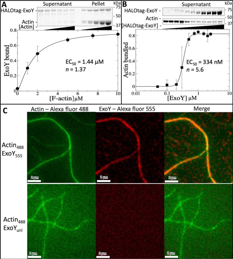
Pseudomonas aeruginosa uses a type III secretion system (T3SS) to inject cytotoxic effector proteins into host cells. The promiscuous nucleotidyl cyclase, exoenzyme Y (ExoY), is one of the most common effectors found in clinical P. aeruginosa isolates. Recent studies have revealed that the nucleotidyl cyclase activity of ExoY is stimulated by actin filaments (F-actin) and that ExoY alters actin cytoskeleton dynamics in vitro, via an unknown mechanism. The actin cytoskeleton plays an important role in numerous key biological processes and is targeted by many pathogens to gain competitive advantages. We utilized total internal reflection fluorescence microscopy, bulk actin assays, and EM to investigate how ExoY impacts actin dynamics. We found that ExoY can directly bundle actin filaments with high affinity, comparable with eukaryotic F-actin-bundling proteins, such as fimbrin. Of note, ExoY enzymatic activity was not required for F-actin bundling. Bundling is known to require multiple actin-binding sites, yet small-angle X-ray scattering experiments revealed that ExoY is a monomer in solution, and previous data suggested that ExoY possesses only one actin-binding site. We therefore hypothesized that ExoY oligomerizes in response to F-actin binding and have used the ExoY structure to construct a dimer-based structural model for the ExoY-F-actin complex. Subsequent mutational analyses suggested that the ExoY oligomerization interface plays a crucial role in mediating F-actin bundling. Our results indicate that ExoY represents a new class of actin-binding proteins that modulate the actin cytoskeleton both directly, via F-actin bundling, and indirectly, via actin-activated nucleotidyl cyclase activity.

Keywords: Pseudomonas aeruginosa; actin; actin bundling; bacterial pathogenesis; bacterial toxin; cytoskeleton; exoenzyme Y (ExoY); host-microbe interactions; host-pathogen interaction; protein-protein interaction; virulence factor.


Conflict of interest statement

The authors declare that they have no conflicts of interest with the contents of this article

Figures

Figure 1.
Figure 1.
ExoY stimulates F-actin bundling. Actin filaments form bundles in the presence of ExoY. A, frame-by-frame time lapse of two actin filaments bundling in the presence of 200 nm ExoY. Actin (1.5 μm) is fluorescently labeled with Alexa Fluor 488. Images were captured via TIRFm. See Fig. S1 for the full movie. Scale bars, 5 μm. B, negatively stained electron micrographs showing that actin filaments bundle in the presence of ExoY (bottom row). Actin filaments in the absence of ExoY (top row) are shown for comparison. Magnification was as indicated; scale bars, 1 μm (×2,900), 200 nm (×11,000), or 50 nm (×30,000). ×11,000 panels for actin and actin + ExoY are included in Fig. 2B for comparative purposes.
Figure 2.
Figure 2.
ExoY enzymatic activity is not required for actin bundling. A, frame-by-frame time lapse of two actin filaments bundling in the presence of catalytically inactive ExoYK81M. Actin (1.5 μm) is fluorescently labeled with Alexa Fluor 488. Images were captured via TIRFm. See Movie S2 for the full movie. Scale bars, 5 μm. B, negatively stained electron micrographs showing that actin filaments bundle in the presence of ExoYK81M (right). Actin filaments in the absence (left) and presence (center) of WT ExoY (panels from Fig. 1B) are shown for comparison. Images were taken at ×11,000 magnification; scale bars, 200 nm. C, micrograph of the WT ExoY-actin bundle in vitreous ice at ×50,000 magnification; scale bar, 50 nm. D, micrograph showing a sample of actin filaments in the presence of catalytically inactive ExoYK81M in vitreous ice. The bundles formed by ExoYK81M do not survive the vitrification process. Magnification was ×50,000; scale bar, 50 nm.
Figure 3.
Figure 3.
ExoY directly cross-links actin filaments within the bundle. ExoY and actin are indistinguishable via SDS-PAGE due to their similar molecular weights; therefore, an N-terminal HaloTag was added to ExoY to increase its size. A, high-speed sedimentation (100,000 × g) demonstrating ExoY actin-binding affinity. Error bars, S.E. of two independent replicates. Data were fit with the Hill equation (curve), which demonstrated that ExoY binds F-actin with positive cooperativity (n = 1.37). B, low-speed sedimentation (10,000 × g) demonstrating ExoY actin-bundling affinity. Error bars, S.E. of two independent replicates. Data were fit with the Hill equation (curve), which demonstrated that ExoY bundles F-actin with positive cooperativity (n = 5.6). C, two-color TIRFm images demonstrating the presence of ExoY within the actin bundle. Actin was labeled with Alexa Fluor 488, and ExoY was labeled with Alexa Fluor 555.
Figure 4.
Figure 4.
Structural model for the ExoY dimer and ExoY-actin bundle. A, SEC-SAXS indicates that ExoY exists as a monomer in solution. The predicted scattering curves calculated from the ExoY crystal structure (orange) match well with experimental data (blue). B, putative ExoY dimer resulting from PISA analysis of the ExoY crystal structure (PDB code 5XNW). Individual subunits are colored in green and magenta, and CA and CB domains are shown in separate shades. Stars indicate the nucleotide-binding pocket. Regions absent in the crystal structure have been joined with dashed lines for ease of viewing. C, the ExoY dimer was docked into the structure of an actin filament (PDB code 3J0S, actin only, individual subunits shown in shades of gray) on the basis of charge complementarity. Asp-25, a residue previously shown to mediate ExoY-actin interaction, is highlighted in cyan. Charge distribution (top panel) was calculated in PyMOL (55). Red, negative charge; white, neutral; blue, positively charged surface. D, model of the ExoY-actin complex. The interaction interface shown in C was superimposed upon the second subunit of the ExoY dimer to generate a 2:2 ExoY:F-actin complex. E, model of the ExoY-actin bundle. Full-length actin filaments (PDB code 3J0S, actin only) were superimposed upon the actin trimers, and a second dimer of ExoY was modeled in following the docking procedure outlined in C.
Figure 5.
Figure 5.
ExoY competes with fimbrin and cofilin for binding to F-actin. A, comparison of cofilin (PDB code 5YU8) (56), fimbrin (PDB code 3BYH) (57), and ExoY-binding sites on F-actin. High-speed (100,000 × g) sedimentation demonstrates that increasing concentrations of HaloTagged ExoY displace 1 μm cofilin or 1 μm SNAP-tagged fimbrin from preassembled actin filaments (1.5 μm). B, representative gels showing that the amount of cofilin and SNAP-tagged fimbrin pelleted decreases as the amount of ExoY increases. C, relative pellet composition. Following SDS-PAGE and Coomassie Blue staining, cofilin or SNAP-tagged fimbrin pellet band densities were quantified and compared with the pellet band density for HaloTagged ExoY. All pellet band densities were normalized to the amount of actin in the pellet as a loading control. Error bars, S.E. of two independent replicates.
Figure 6.
Figure 6.
Mutations at the putative dimer interface alter ExoY-actin bundling. Characterization of the E175S/E177S (A), H104S (B), and D156S (C) mutations. The top row of each panel shows a schematic of the ExoY dimer with key residues indicated, colored as in Fig. 4. The middle row depicts the quantification of mutant construct bundling efficiency (open circles, dashed line) compared with WT ExoY (closed squares, solid line). Data were fit to the Hill equation. The bottom row shows the quantification of high-speed sedimentation assays demonstrating construct (open circle, dashed line) binding to F-actin compared with WT ExoY (closed squares, solid line). Error bars, S.E. of two independent replicates. For low-speed assays, the concentration of ExoY ranged from 0 to 2.5 μm (WT), from 0 to 500 nm (E175S/E177S), and from 0 to 10 μm (H104S and D156S), whereas the concentration of actin was kept constant at 1.5 μm. For the high-speed assays, 1.5 μm ExoY was tested against a range of actin concentrations spanning 0–10 μm. Actin concentrations denote G-actin concentration prior to polymerization. It was assumed that actin was fully polymerized prior to the start of the assay.

References

    1. Micek S. T., Wunderink R. G., Kollef M. H., Chen C., Rello J., Chastre J., Antonelli M., Welte T., Clair B., Ostermann H., Calbo E., Torres A., Menichetti F., Schramm G. E., and Menon V. (2015) An international multicenter retrospective study of Pseudomonas aeruginosa nosocomial pneumonia: impact of multidrug resistance. Crit. Care 19, 219 10.1186/s13054-015-0926-5 - DOI - PMC - PubMed
    1. Hauser A. R. (2009) The type III secretion system of Pseudomonas aeruginosa: infection by injection. Nat. Rev. Microbiol. 7, 654–665 10.1038/nrmicro2199 - DOI - PMC - PubMed
    1. Feltman H., Schulert G., Khan S., Jain M., Peterson L., and Hauser A. R. (2001) Prevalence of type III secretion genes in clinical and environmental isolates of Pseudomonas aeruginosa. Microbiology 147, 2659–2669 10.1099/00221287-147-10-2659 - DOI - PubMed
    1. Sato H., Frank D. W., Hillard C. J., Feix J. B., Pankhaniya R. R., Moriyama K., Finck-Barbançon V., Buchaklian A., Lei M., Long R. M., Wiener-Kronish J., and Sawa T. (2003) The mechanism of action of the Pseudomonas aeruginosa-encoded type III cytotoxin, ExoU. EMBO J. 22, 2959–2969 10.1093/emboj/cdg290 - DOI - PMC - PubMed
    1. Belyy A., Santecchia I., Renault L., Bourigault B., Ladant D., and Mechold U. (2018) The extreme C terminus of the Pseudomonas aeruginosa effector ExoY is crucial for binding to its eukaryotic activator, F-actin. J. Biol. Chem. 293, 19785–19796 10.1074/jbc.RA118.003784 - DOI - PMC - PubMed

Publication types

MeSH terms

LinkOut - more resources