

Necrostatin-1 Alleviates Bleomycin-Induced Pulmonary Fibrosis and Extracellular Matrix Expression in Interstitial Pulmonary Fibrosis
- PMID: 32019905
- PMCID: PMC7020761
- DOI: 10.12659/MSM.919739
Necrostatin-1 Alleviates Bleomycin-Induced Pulmonary Fibrosis and Extracellular Matrix Expression in Interstitial Pulmonary Fibrosis
Abstract
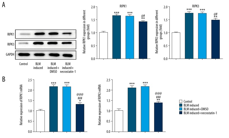
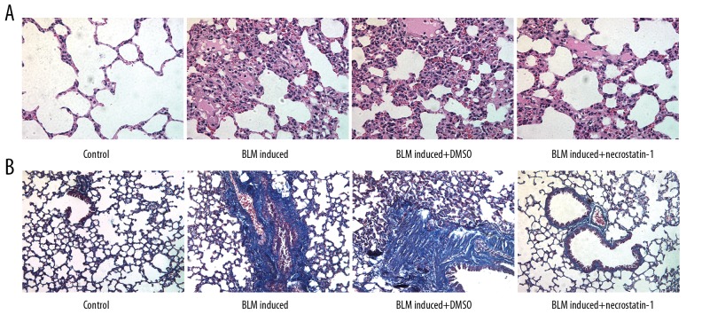
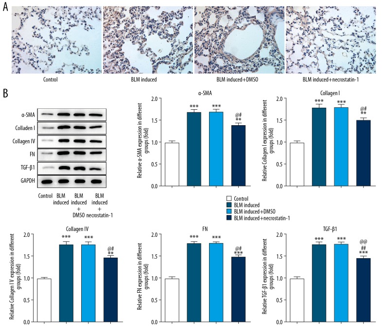
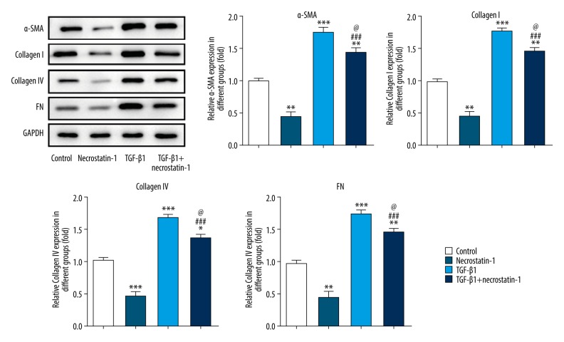
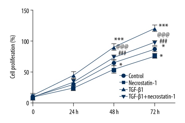
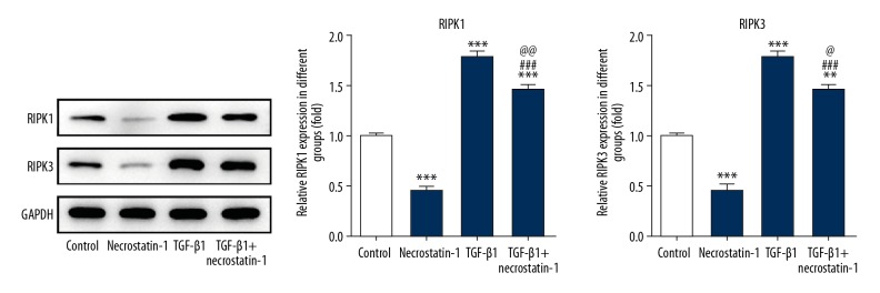
BACKGROUND Interstitial pulmonary fibrosis (IPF) is harmful for patients' life and health. The effective treatment of IPF is lacking because of unclear pathogenesis. Necrostatin-1 has protective effects on lung injury and can suppress the fibrosis development. I this study we investigated whether necrostatin-1 could decrease the proliferation of pulmonary fibroblasts, pulmonary fibrosis and expression of extracellular matrix (ECM) in IPF. MATERIAL AND METHODS The IPF mice model was conducted by intra-tracheal injection of bleomycin (BLM) (2 mg/kg) for C57BL/6N mice. Necrostatin-1 treatment was performed with 1 mg/kg necrostatin-1 by an intravenous injection for C57BL/6N mice. Lung tissue structures and collagen deposition were observed by hematoxylin and eosin staining and Masson staining. IPF in vitro model was constructed by MRC-5 cells induced by transforming growth factor beta 1 (TGF-ß1). And, 20 μM necrostatin-1 was used to treat the TGF-ß1 induced MRC-5 cells. Cell Counting Kit-8 (CCK-8) assay detected the viability of MRC-5 cells. The expression of receptor-interacting protein kinase-1 and -3 (RIPK1 and RIPK3), alpha smooth muscle actin (alpha-SMA), collagen IV, collagen I, fibronectin (FN), and transforming growth factor-ß (TGF-ß) in lung tissues and MRC-5 cells was measured by western blot analysis. The alpha-SMA expression in lung tissues was also analyzed by immunohistochemistry. RESULTS The expression of RIPK1 and RIPK3 in lung tissues of BLM induced mice was increased. The degree of pulmonary fibrosis and expression of alpha-SMA, collagen IV, collagen I, FN, and TGF-ß in lung tissues of BLM induced mice was enhanced. The proliferation of MRC-5 cells was increased when MRC-5 cells were induced by TGF-ß. The expression of RIPK1, RIPK3, alpha-SMA, collagen IV, collagen I, and FN was increased in TGF-ß induced MRC-5 cells. And, necrostatin-1 could effectively reverse the changes of pulmonary fibrosis, RIPK1, RIPK3, and ECM in vivo and in vitro experiments. CONCLUSIONS Necrostatin-1 attenuated pulmonary fibrosis in lung tissues of BLM induced mice and inhibited the fibroblast proliferation. And, necrostatin-1 also decreased the expression of RIPK1, RIPK3, and ECM in lung tissues of BLM induced mice and TGF-ß induced fibroblasts. Necrostatin-1 could be a new effective drug for the treatment of IPF.
Conflict of interest statement
None.
Figures
Similar articles
-
[Digoxin alleviates pulmonary fibrosis by regulating phosphatidylinositol-3-kinase/Akt signaling through inhibiting the activation of fibroblast: an in vivo and in vitro experiment].Zhonghua Wei Zhong Bing Ji Jiu Yi Xue. 2022 Nov;34(11):1161-1166. doi: 10.3760/cma.j.cn121430-20220628-00508. Zhonghua Wei Zhong Bing Ji Jiu Yi Xue. 2022. PMID: 36567559 Chinese.
-
Sulforaphane attenuates pulmonary fibrosis by inhibiting the epithelial-mesenchymal transition.BMC Pharmacol Toxicol. 2018 Apr 2;19(1):13. doi: 10.1186/s40360-018-0204-7. BMC Pharmacol Toxicol. 2018. PMID: 29609658 Free PMC article.
-
RS4651 suppresses lung fibroblast activation via the TGF-β1/SMAD signalling pathway.Eur J Pharmacol. 2021 Jul 15;903:174135. doi: 10.1016/j.ejphar.2021.174135. Epub 2021 May 1. Eur J Pharmacol. 2021. PMID: 33940030
-
The extracellular matrix and mechanotransduction in pulmonary fibrosis.Int J Biochem Cell Biol. 2020 Sep;126:105802. doi: 10.1016/j.biocel.2020.105802. Epub 2020 Jul 12. Int J Biochem Cell Biol. 2020. PMID: 32668329 Review.
-
PTX3 Regulation of Inflammation, Hemostatic Response, Tissue Repair, and Resolution of Fibrosis Favors a Role in Limiting Idiopathic Pulmonary Fibrosis.Front Immunol. 2021 Jun 21;12:676702. doi: 10.3389/fimmu.2021.676702. eCollection 2021. Front Immunol. 2021. PMID: 34276664 Free PMC article. Review.
Cited by
-
Recent Advances (2015-2020) in Drug Discovery for Attenuation of Pulmonary Fibrosis and COPD.Molecules. 2023 Apr 24;28(9):3674. doi: 10.3390/molecules28093674. Molecules. 2023. PMID: 37175084 Free PMC article. Review.
-
PANoptosis: Mechanism and Role in Pulmonary Diseases.Int J Mol Sci. 2023 Oct 19;24(20):15343. doi: 10.3390/ijms242015343. Int J Mol Sci. 2023. PMID: 37895022 Free PMC article. Review.
-
[Research Progress of Anti-lung Cancer Drug-related Interstitial Lung Disease].Zhongguo Fei Ai Za Zhi. 2025 Apr 20;28(4):309-318. doi: 10.3779/j.issn.1009-3419.2025.106.11. Zhongguo Fei Ai Za Zhi. 2025. PMID: 40404479 Free PMC article. Review. Chinese.
-
Exploring the Mechanism Whereby Sinensetin Delays the Progression of Pulmonary Fibrosis Based on Network Pharmacology and Pulmonary Fibrosis Models.Front Pharmacol. 2021 Jun 18;12:693061. doi: 10.3389/fphar.2021.693061. eCollection 2021. Front Pharmacol. 2021. PMID: 34220517 Free PMC article.
-
RIPK3 Expression in Fibroblasts in an in vivo and in vitro Skin Wound Model: A Controversial Result.Acta Naturae. 2023 Oct-Dec;15(4):65-74. doi: 10.32607/actanaturae.25452. Acta Naturae. 2023. PMID: 38234604 Free PMC article.
References
-
- Mari PV, Jones MG, Richeldi L. Contemporary concise review 2018: Interstitial lung disease. Respirology. 2019;24:809–16. - PubMed
-
- Chinese Society of Respiratory Diseases. Draft Guidelines for the diagnosis and treatment of idiopathic pulmonary (interstitial) fibrosis. Chin J Tuberc Respir Dis. 2002;25:387–89.
-
- Interstitial Pulmonary Disease Committee RS, Chinese Medical Association. Chinese expert consensus on diagnosis and treatment of idiopathic pulmonary fibrosis. Chin J Tuberc Respir Dis. 2016;39:427–32.
-
- Raghu G, Rochwerg B, Zhang Y, et al. An Official ATS/ERS/JRS/ALAT Clinical practice guideline: Treatment of idiopathic pulmonary fibrosis. An update of the 2011 clinical practice guideline. Am J Respir Crit Care Med. 2015;192:e3–19. - PubMed
MeSH terms
Substances
LinkOut - more resources
Full Text Sources
Medical
Miscellaneous
