. 2020 Feb 4;11(1):694.
doi: 10.1038/s41467-020-14340-x.

Sustained treatment of retinal vascular diseases with self-aggregating sunitinib microparticles

Affiliations

Sustained treatment of retinal vascular diseases with self-aggregating sunitinib microparticles

Hiroki Tsujinaka et al. Nat Commun. .

Abstract

Neovascular age-related macular degeneration and diabetic retinopathy are prevalent causes of vision loss requiring frequent intravitreous injections of VEGF-neutralizing proteins, and under-treatment is common and problematic. Here we report incorporation of sunitinib, a tyrosine kinase inhibitor that blocks VEGF receptors, into a non-inflammatory biodegradable polymer to generate sunitinib microparticles specially formulated to self-aggregate into a depot. A single intravitreous injection of sunitinib microparticles potently suppresses choroidal neovascularization in mice for six months and in another model, blocks VEGF-induced leukostasis and retinal nonperfusion, which are associated with diabetic retinopathy progression. After intravitreous injection in rabbits, sunitinib microparticles self-aggregate into a depot that remains localized and maintains therapeutic levels of sunitinib in retinal pigmented epithelium/choroid and retina for more than six months. There is no intraocular inflammation or retinal toxicity. Intravitreous injection of sunitinib microparticles provides a promising approach to achieve sustained suppression of VEGF signaling and improve outcomes in patients with retinal vascular diseases.


Conflict of interest statement

J.H., J.F. and P.A.C. are founders of Graybug Vision which has a commercial interest in sunitinib microparticles, and they have equity in Graybug and receive remuneration for consultation. Johns Hopkins University has patents on which J.H. and J.F. are authors that are licensed to Graybug Vision. All of these relationships are being managed by the Johns Hopkins University conflict of interest committee. Y.Y., D.M., D.Ca., W.Y. and M.Y. are employees of Graybug Vision. J.K., W.P., J.Z.Z. and J.L.C. are former employees of Graybug Vision. The remaining authors declare no competing interests.

Figures
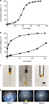
Fig. 1
Fig. 1. Characterization of sunitinib MPs and degradation of sunitinib MP depot in the vitreous of minipigs.
a Sunitinib MPs used in rabbit and minipig experiments release drug in a sustained manner for nearly 3 months in vitro at 37 °C under sink conditions. Circles are the means for two independent experiments. b Sunitinib drug suspension (open squares) was rapidly eluted and dissolved in release medium in vitro, while MPs containing an equivalent amount of sunitinib (closed squares) achieved a much more sustained release over time. c An injection of 50 µl sunitinib MP suspension in a hyaluronate solution at 37 °C leads to the formation of a solid depot that can be isolated using a pair of forceps. d Wide-field fundus photographs showing the reduction of depot size over time (Day 8, 43, and 92 post dose) in vitreous after an intravitreous injection of MP suspension containing 1 mg sunitinib in minipigs. The yellow spot near the bottom of each photo indicates the sunitinib MP depot. Source data are provided as a Source Data file.
Fig. 2
Fig. 2. Effect of sunitinib microparticles in a mouse model of type 2 choroidal neovascularization.
C57BL/6 mice received a 1 µl intravitreous injection of sunitinib (Suni) microparticles (MP) containing 10 or 1 µg of sunitinib in one eye and an equivalent amount of empty MP in the fellow eye, or they received an injection of 0.5 or 0.1 µg of free sunitinib in one eye and PBS in the fellow eye, or 40 µg of aflibercept in one eye and PBS in the fellow eye. At various time points after injection ranging from 1 to 24 weeks, the mice had rupture of Bruch’s membrane by laser photocoagulation at 3 locations in each eye. One week after laser, mice were euthanized and choroidal flat mounts were stained with FITC-labeled Griffonia Simplicifolia Agglutinin (GSA) lectin and the area of choroidal neovascularization (CNV) was measured at Bruch’s membrane rupture sites by an investigator masked with regard to treatment. The three values from each eye were averaged to give a single experimental value. The mean (±SEM) area of CNV was significantly less in eyes injected with MP containing 10 µg sunitinib compared with fellow eyes injected with empty MP at each time point through 24 weeks (a). Eyes injected with MP containing 1 µg sunitinib showed a significant reduction in mean area of CNV compared with corresponding controls at 9 and 15 weeks, but not 20 and 24 weeks (b). Eyes injected with 0.5 or 0.1 µg of free sunitinib showed no significant reduction in area of CNV compared to fellow eyes injected with PBS at 1 week (c). Eyes injected with 40 µg of aflibercept showed a significant reduction in mean CNV area compared with fellow eye controls at 1 week, but not 9 or 15 weeks (d). Number of animals used in each group are shown in the graph or below the graph. *p < 0.05; **p < 0.01, ***p < 0.001 by Mann–Whitney compared with fellow eye control at that time point. Bar = 100 µm. Source data are provided as a Source Data file.
Fig. 3
Fig. 3. Subconjunctival injection of sunitinib MPs suppresses type 2 choroidal NV.
C57BL/6 mice received a 2 µl subconjunctival injection of microparticles (MP) containing 20 or 2 µg of sunitinib (Suni) or empty MP in one eye and no injection in the fellow eye. At 1 week after injection, Bruch’s membrane was ruptured by laser photocoagulation at three locations in each eye. One week after laser, mice were euthanized and choroidal flat mounts were stained with FITC-labeled GSA lectin and the area of choroidal neovascularization (CNV) was measured at Bruch’s membrane rupture sites by an investigator masked with regard to treatment. The three values from each eye were averaged to give a single experimental value. The mean (±SEM) area of CNV was significantly less in eyes injected with MP containing 20 µg (a) or 2 µg (b) sunitinib vs. those injected with empty MP, but there was no significant difference between fellow eyes for each group. *p < 0.05 for difference from all other groups by Kruskal Wallis test followed by Dunn’s test. Bar = 100 µm. Source data are provided as a Source Data file.
Fig. 4
Fig. 4. Sunitinib MPs suppress murine type 3 choroidal NV substantially longer than aflibercept.
At P14, rho/VEGF mice had intravitreous injection of MP containing 10 µg sunitinib (Suni MP) in one eye and empty MPs in the other eye or 40 µg of aflibercept in one eye and PBS in the other eye. At P21, P28, P35, or P42, mice were euthanized and retinal flat mounts were stained with FITC-labeled GSA lectin. The total area of subretinal NV per retina was measured by image analysis. Compared with empty MP-injected eyes, those injected with Suni MPs had significantly lower mean (±SEM) area of NV per retina at each time point (a). Compared with PBS-injected eyes, those injected with aflibercept had significantly lower mean (±SEM) area of NV per retina at P21, but not P28, P35, or P42 (b). At P28, rho/VEGF mice had fluorescein angiography showing severe leakage resulting in large collections of extravascular fluorescein (c top row). One eye was injected with MP containing 10 µg of sunitinib or 40 µg of aflibercept and the other with empty MPs or PBS and after 1 week repeat fluorescein angiography showed less leakage in sunitinib MP-injected eyes, but not empty MP-injected eyes (c bottom row, scale bar = 100 µm). Vitreous samples were obtained and the mean (±SEM) concentration of vitreous albumin measured by ELISA was significantly less in Suni MP-injected eyes vs. empty MP-injected eyes and similar to untreated control eyes (d). Mean vitreous albumin was also significantly less in aflibercept-injected eyes vs. PBS-injected eyes (d). The experiment described in (c) and (d) was repeated in P28 rho/VEGF mice using a different outcome measure, leakage of intravascular Evans Blue dye into the retina as described in Methods. e The mean (±SEM) concentration of Evans Blue dye in the retina was significantly less in eyes injected with sunitinib MP compared with those injected with empty MP. Number of animals used in each group are shown in the graph or below the graph. *p < 0.05; **p < 0.01; ***p < 0.001 by Mann Whitney from corresponding control. Bar = 100 µm. Source data are provided as a Source Data file.
Fig. 5
Fig. 5. Intravitreous injection of sunitinib MPs in mice with type 3 CNV reduces photoreceptor cell death.
Rho/VEGF mice were given an intravitreous injection of 10 µg of sunitinib microparticles (Suni MP) in one eye and 10 µg of empty MP in the other eye or 40 µg of aflibercept in one eye and PBS in the fellow eye. At P42, mice (n = 7 for each group) were euthanized and serial frozen ocular sections were cut from the superior pole of the eye (S0) to the inferior pole (I0) and sections 25% (S1 and I1), 50% (S2 and I2), and 75% (S3 and I3) of the distance between each pole and the optic nerve (ON) were stained with hematoxylin and outer nuclear layer (ONL) thickness was measured by image analysis by a masked investigator. The ONL of sections from the S2 location of Suni MP-injected eyes appeared thicker than those from empty MP-injected eyes, but those from aflibercept-injected eyes appeared similar to those from PBS-injected eyes (a scale bar = 100 µm). The mean (±SEM) ONL thickness was significantly greater at three of six locations in Suni MP-injected eyes compared with empty MP-injected eyes, but there was no difference between aflibercept- and PBS-injected eyes (b). At P49, mice (n = 5 for each group) were euthanized and immunoblots of retinal homogenates from Suni MP-injected eyes showed prominent bands for rhodopsin kinase (GRK-1) (c). Denistometry showed that the mean (±SEM) GRK-1/Actin ratio was significantly greater in retinas from Suni MP-injected eyes compared with empty MP-injected fellow eyes, but not aflibercept-injected eyes vs. PBS-injected fellow eyes (d). *p < 0.05 by Mann Whitney for difference from fellow eye empty MP control. Source data are provided as a Source Data file.
Fig. 6
Fig. 6. Sunitinib MP reduce leukostasis and improve retinal perfusion in rho/VEGF mice.
Rho/VEGF mice were given an intravitreous injection of 10 µg of Suni MP or 40 µg of aflibercept in one eye and 10 µg of Empty MP or PBS in the other eye. After 1 week, mice were perfused through the left ventricle with PBS to remove erythrocytes and leukocytes and then perfused with FITC-concanavalin A. Examination of retinal flat mounts by fluorescence microscopy showed a significant reduction in mean number of adherent intravascular leukocytes per retina in Suni MP-injected eyes compared with Empty MP-injected fellow eyes and in aflibercept-injected eyes compared with PBS-injected fellow eyes (a scale bar = 100 µm; *p < 0.001 by Mann Whitney for difference from corresponding control). The experiment was repeated but leukostasis was visualized by immunohistochemical staining with anti-CD45 which showed a significant reduction in leukocytes within retinal vessels in eyes injected with Suni MPs vs. those injected with empty MPs (b scale bar = 100 µm; *p < 0.001 by Mann Whitney for difference from corresponding fellow eye control). Rho/VEGF mice had fluorescein angiography at baseline and then were given an intravitreous injection of 10 µg of Suni MP in one eye and 10 µg of Empty MP in the other eye. Some areas of retinal nonperfusion at baseline showed improved perfusion 1 week after injection of Suni MP, but this was not seen in Empty MP-injected eyes (c scale bar = 250 µm). Source data are provided as a Source Data file.
Fig. 7
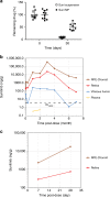
Fig. 7. Pharmacokinetic profiles of sunitinib MPs.
a C57BL/6 mice were given a 1 µl intravitreous injection of either MPs containing 1 µg sunitinib (closed bar) or an equivalent dose of sunitinib drug suspension (open bar) in both eyes. Immediately after injection or at 30 days after injection, 5 mice from each condition were euthanized, and the amount of sunitinib remaining in the eyes was evaluated by a validated extraction and the LC–MS method and presented as a percentage of the initial dose. b A 50 µl suspension of sunitinib MPs containing 1 mg sunitinib was administered to both eyes of pigmented New Zealand rabbits. After 500 μL of blood was collected, animals were euthanized and the eyes were enucleated immediately after sacrifice. Central region RPE-choroid, retina, and vitreous humor in one eye of each animal were collected for pharmacokinetic ocular tissue analysis at 24 h, Days 3 and 10, and Months 1–4, 6, and 7. N = 4 biologically independent samples for time points through 4 month, n = 2 for 6 months, and n = 1 for 7 months. c A 200 µl suspension of sunitinib MPs containing 2 mg sunitinib was administered to both eyes of pigmented New Zealand rabbits. Central region RPE-choroid and retina in one eye of each animal were collected Days 7 and 28 (n = 3 biologically independent samples for each tissue at each time point). Sunitinib levels in plasma and ocular tissues were evaluated by LC–MS. Source data are provided as a Source Data file.

Similar articles

Cited by

References

    1. Klein R, Klein BE, Tomany SC, Meuer SM, Huang GH. Ten-year incidence and progression of age-related macular degeneration: the Beaver Dam Eye study. Ophthalmology. 2002;109:1767–1779. doi: 10.1016/S0161-6420(02)01146-6. - DOI - PubMed
    1. Grunwald JE, et al. Risk of geographic atrophy in the comparison of age-related macular degeneration treatments trials. Ophthalmology. 2012;121:150–161. doi: 10.1016/j.ophtha.2013.08.015. - DOI - PMC - PubMed
    1. Chew EY, et al. Ten-year follow-up of age-related macular degeneration in the age-related eye disease study. JAMA Ophthalmol. 2014;132:272–277. doi: 10.1001/jamaophthalmol.2013.6636. - DOI - PMC - PubMed
    1. Kwak N, Okamoto N, Wood JM, Campochiaro PA. VEGF is an important stimulator in a model of choroidal neovascularization. Invest. Ophthalmol. Vis. Sci. 2000;41:3158–3164. - PubMed
    1. Kryzstolik MG, et al. Prevention of experimental choroidal neovascularization with intravitreal anti-vascular endothelial growth factor antibody fragment. Arch. Ophthalmol. 2002;120:338–346. doi: 10.1001/archopht.120.3.338. - DOI - PubMed

Publication types

MeSH terms

Substances